8QY6

Structure of interleukin 6 (gp130 P496L mutant).

  • Classification: IMMUNE SYSTEM
  • Organism(s): Mus musculus, Homo sapiens
  • Expression System: Trichoplusia ni
  • Mutation(s): Yes 

  • Deposited: 2023-10-25 Released: 2024-02-21 
  • Deposition Author(s): Gardner, S., Bubeck, D., Jin, Y.
  • Funding Organization(s): Wellcome Trust, European Research Council (ERC), Engineering and Physical Sciences Research Council, Biotechnology and Biological Sciences Research Council (BBSRC)

Experimental Data Snapshot

  • Method: ELECTRON MICROSCOPY
  • Resolution: 3.16 Å
  • Aggregation State: PARTICLE 
  • Reconstruction Method: SINGLE PARTICLE 

wwPDB Validation   3D Report Full Report


This is version 1.1 of the entry. See complete history


Literature

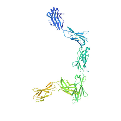
Structural insights into IL-11-mediated signalling and human IL6ST variant-associated immunodeficiency.

Gardner, S.Jin, Y.Fyfe, P.K.Voisin, T.B.Bellon, J.S.Pohler, E.Piehler, J.Moraga, I.Bubeck, D.

(2024) Nat Commun 15: 2071-2071

  • DOI: https://doi.org/10.1038/s41467-024-46235-6
  • Primary Citation of Related Structures:  
    8QY4, 8QY5, 8QY6

  • PubMed Abstract: 

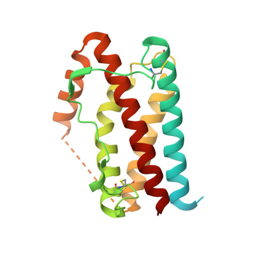
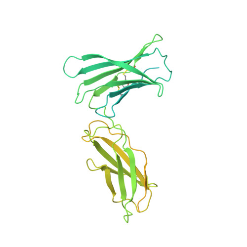
    IL-11 and IL-6 activate signalling via assembly of the cell surface receptor gp130; however, it is unclear how signals are transmitted across the membrane to instruct cellular responses. Here we solve the cryoEM structure of the IL-11 receptor recognition complex to discover how differences in gp130-binding interfaces may drive signalling outcomes. We explore how mutations in the IL6ST gene encoding for gp130, which cause severe immune deficiencies in humans, impair signalling without blocking cytokine binding. We use cryoEM to solve structures of both IL-11 and IL-6 complexes with a mutant form of gp130 associated with human disease. Together with molecular dynamics simulations, we show that the disease-associated variant led to an increase in flexibility including motion within the cytokine-binding core and increased distance between extracellular domains. However, these distances are minimized as the transmembrane helix exits the membrane, suggesting a stringency in geometry for signalling and dimmer switch mode of action.


  • Organizational Affiliation

    Department of Life Sciences, Sir Ernst Chain Building, Imperial College London, London, SW7 2AZ, UK.


Macromolecules
Find similar proteins by:  (by identity cutoff)  |  3D Structure
Entity ID: 1
MoleculeChains Sequence LengthOrganismDetailsImage
Interleukin-6 receptor subunit betaA,
F [auth D]
917Mus musculusMutation(s): 1 
Gene Names: Il6st
UniProt & NIH Common Fund Data Resources
Find proteins for Q00560 (Mus musculus)
Explore Q00560 
Go to UniProtKB:  Q00560
IMPC:  MGI:96560
Entity Groups  
Sequence Clusters30% Identity50% Identity70% Identity90% Identity95% Identity100% Identity
UniProt GroupQ00560
Glycosylation
Glycosylation Sites: 6Go to GlyGen: Q00560-1
Sequence Annotations
Expand
  • Reference Sequence
Find similar proteins by:  (by identity cutoff)  |  3D Structure
Entity ID: 2
MoleculeChains Sequence LengthOrganismDetailsImage
Interleukin-6B,
D [auth E]
212Homo sapiensMutation(s): 0 
Gene Names: IL6
UniProt & NIH Common Fund Data Resources
Find proteins for P05231 (Homo sapiens)
Explore P05231 
Go to UniProtKB:  P05231
PHAROS:  P05231
GTEx:  ENSG00000136244 
Entity Groups  
Sequence Clusters30% Identity50% Identity70% Identity90% Identity95% Identity100% Identity
UniProt GroupP05231
Sequence Annotations
Expand
  • Reference Sequence
Find similar proteins by:  (by identity cutoff)  |  3D Structure
Entity ID: 3
MoleculeChains Sequence LengthOrganismDetailsImage
Interleukin-6 receptor subunit alphaC,
E [auth F]
468Homo sapiensMutation(s): 0 
Gene Names: IL6R
UniProt & NIH Common Fund Data Resources
Find proteins for P08887 (Homo sapiens)
Explore P08887 
Go to UniProtKB:  P08887
PHAROS:  P08887
GTEx:  ENSG00000160712 
Entity Groups  
Sequence Clusters30% Identity50% Identity70% Identity90% Identity95% Identity100% Identity
UniProt GroupP08887
Sequence Annotations
Expand
  • Reference Sequence
Oligosaccharides

Help

Entity ID: 4
MoleculeChains Length2D Diagram Glycosylation3D Interactions
2-acetamido-2-deoxy-beta-D-glucopyranose-(1-4)-2-acetamido-2-deoxy-beta-D-glucopyranose
G, H, I, J, K
G, H, I, J, K, L, M, N, O, P
2N-Glycosylation
Glycosylation Resources
GlyTouCan:  G42666HT
GlyCosmos:  G42666HT
GlyGen:  G42666HT
Small Molecules
Ligands 1 Unique
IDChains Name / Formula / InChI Key2D Diagram3D Interactions
NAG
Query on NAG

Download Ideal Coordinates CCD File 
Q [auth A],
R [auth D]
2-acetamido-2-deoxy-beta-D-glucopyranose
C8 H15 N O6
OVRNDRQMDRJTHS-FMDGEEDCSA-N
Experimental Data & Validation

Experimental Data

  • Method: ELECTRON MICROSCOPY
  • Resolution: 3.16 Å
  • Aggregation State: PARTICLE 
  • Reconstruction Method: SINGLE PARTICLE 
EM Software:
TaskSoftware PackageVersion
MODEL REFINEMENTPHENIX1.21rc1_4933

Structure Validation

View Full Validation Report


Entry History & Funding Information

Deposition Data


Funding OrganizationLocationGrant Number
Wellcome TrustUnited Kingdom202323/Z/16
European Research Council (ERC)European UnionC-206-STG
Engineering and Physical Sciences Research CouncilUnited KingdomEP/X035603/1
Biotechnology and Biological Sciences Research Council (BBSRC)United KingdomBB/M011178/1

Revision History  (Full details and data files)

  • Version 1.0: 2024-02-21
    Type: Initial release
  • Version 1.1: 2024-09-04
    Changes: Data collection, Database references