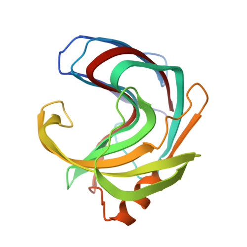
Bimodal substrate binding in the active site of the glycosidase BcX.
Saberi, M., Chikunova, A., Ben Bdira, F., Cramer-Blok, A., Timmer, M., Voskamp, P., Ubbink, M.(2024) FEBS J 291: 4222-4239
- PubMed: 39185686 Search on PubMed
- DOI: https://doi.org/10.1111/febs.17251
- Primary Citation Related Structures:
8QXY, 8QXZ, 8QY0, 8QY1, 8QY2, 8QY3, 8R85, 8R86 - PubMed Abstract:
Bacillus circulans xylanase (BcX) from the glycoside hydrolase family 11 degrades xylan through a retaining, double-displacement mechanism. The enzyme is thought to hydrolyze glycosidic bonds in a processive manner and has a large, active site cleft, with six subsites allowing the binding of six xylose units. Such an active site architecture suggests that oligomeric xylose substrates can bind in multiple ways. In the crystal structure of the catalytically inactive variant BcX E78Q, the substrate xylotriose is observed in the active site, as well as bound to the known secondary binding site and a third site on the protein surface. Nuclear magnetic resonance (NMR) titrations with xylose oligomers of different lengths yield nonlinear chemical shift trajectories for active site nuclei resonances, indicative of multiple binding orientations for these substrates for which binding and dissociation are in fast exchange on the NMR timescale, exchanging on the micro- to millisecond timescale. Active site binding can be modeled with a 2 : 1 model with dissociation constants in the low and high millimolar range. Extensive mutagenesis of active site residues indicates that tight binding occurs in the glycon binding site and is stabilized by Trp9 and the thumb region. Mutations F125A and W71A lead to large structural rearrangements. Binding at the glycon site is sensed throughout the active site, whereas the weak binding mostly affects the aglycon site. The interactions with the two active site locations are largely independent of each other and of binding at the secondary binding site.
- Leiden Institute of Chemistry, Leiden University, The Netherlands.
Organizational Affiliation: