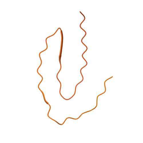
Cryo-EM observation of the amyloid key structure of polymorphic TDP-43 amyloid fibrils.
Sharma, K., Stockert, F., Shenoy, J., Berbon, M., Abdul-Shukkoor, M.B., Habenstein, B., Loquet, A., Schmidt, M., Fandrich, M.(2024) Nat Commun 15: 486-486
- PubMed: 38212334
- DOI: https://doi.org/10.1038/s41467-023-44489-0
- Primary Citation of Related Structures:
8QX9, 8QXA, 8QXB - PubMed Abstract:
The transactive response DNA-binding protein-43 (TDP-43) is a multi-facet protein involved in phase separation, RNA-binding, and alternative splicing. In the context of neurodegenerative diseases, abnormal aggregation of TDP-43 has been linked to amyotrophic lateral sclerosis and frontotemporal lobar degeneration through the aggregation of its C-terminal domain. Here, we report a cryo-electron microscopy (cryo-EM)-based structural characterization of TDP-43 fibrils obtained from the full-length protein. We find that the fibrils are polymorphic and contain three different amyloid structures. The structures differ in the number and relative orientation of the protofilaments, although they share a similar fold containing an amyloid key motif. The observed fibril structures differ from previously described conformations of TDP-43 fibrils and help to better understand the structural landscape of the amyloid fibril structures derived from this protein.
Organizational Affiliation:
Institute of Protein Biochemistry, Ulm University, 89081, Ulm, Germany. kartikay.sharma@uni-ulm.de.


