8QT6 | pdb_00008qt6

Cryo-EM structure of Streptococcus pneumoniae NADPH oxidase


Experimental Data Snapshot

  • Method: ELECTRON MICROSCOPY
  • Resolution: 2.29 Å
  • Aggregation State: PARTICLE 
  • Reconstruction Method: SINGLE PARTICLE 

Starting Model: in silico
View more details

wwPDB Validation 3D Report Full Report

Validation slider image for 8QT6

This is version 1.2 of the entry. See complete history

Literature

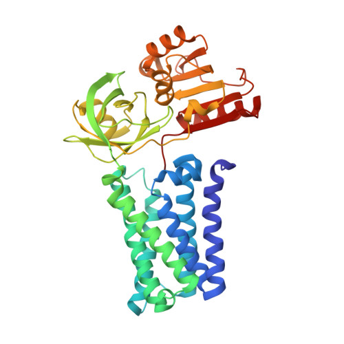
Structural and mechanistic insights into Streptococcus pneumoniae NADPH oxidase.

Dubach, V.R.A.San Segundo-Acosta, P.Murphy, B.J.

(2024) Nat Struct Mol Biol 31: 1769-1777

  • DOI: https://doi.org/10.1038/s41594-024-01348-w
  • Primary Citation Related Structures: 
    8QT6, 8QT7, 8QT9, 8QTA

  • PubMed Abstract: 

    Nicotinamide adenine dinucleotide phosphate (NADPH) oxidases (NOXs) have a major role in the physiology of eukaryotic cells by mediating reactive oxygen species production. Evolutionarily distant proteins with the NOX catalytic core have been found in bacteria, including Streptococcus pneumoniae NOX (SpNOX), which is proposed as a model for studying NOXs because of its high activity and stability in detergent micelles. We present here cryo-electron microscopy structures of substrate-free and nicotinamide adenine dinucleotide (NADH)-bound SpNOX and of NADPH-bound wild-type and F397A SpNOX under turnover conditions. These high-resolution structures provide insights into the electron-transfer pathway and reveal a hydride-transfer mechanism regulated by the displacement of F397. We conducted structure-guided mutagenesis and biochemical analyses that explain the absence of substrate specificity toward NADPH and suggest the mechanism behind constitutive activity. Our study presents the structural basis underlying SpNOX enzymatic activity and sheds light on its potential in vivo function.


  • Organizational Affiliation
    • Redox and Metalloprotein Research Group, Max Planck Institute of Biophysics, Frankfurt am Main, Germany.

Macromolecule Content 

  • Total Structure Weight: 48.08 kDa 
  • Atom Count: 3,466 
  • Modeled Residue Count: 399 
  • Deposited Residue Count: 399 
  • Unique protein chains: 1

Macromolecules

Find similar proteins by:|  3D Structure
Entity ID: 1
MoleculeChains  Sequence LengthOrganismDetailsImage
FAD-binding FR-type domain-containing protein399Streptococcus pneumoniaeMutation(s): 0 
Gene Names: spr0531
Membrane Entity: Yes 
UniProt
Find proteins for Q8CZ28 (Streptococcus pneumoniae (strain ATCC BAA-255 / R6))
Explore Q8CZ28 
Go to UniProtKB:  Q8CZ28
Entity Groups
Sequence Clusters30% Identity50% Identity70% Identity90% Identity95% Identity100% Identity
UniProt GroupQ8CZ28
Sequence Annotations
Expand
Reference Sequence

Experimental Data & Validation

Experimental Data

  • Method: ELECTRON MICROSCOPY
  • Resolution: 2.29 Å
  • Aggregation State: PARTICLE 
  • Reconstruction Method: SINGLE PARTICLE 
EM Software:
TaskSoftware PackageVersion
MODEL REFINEMENTPHENIX
MODEL REFINEMENTCoot
RECONSTRUCTIONcryoSPARC

Structure Validation

View Full Validation Report


Entry History 

& Funding Information

Deposition Data


Funding OrganizationLocationGrant Number
Max Planck SocietyGermany--

Revision History  (Full details and data files)

  • Version 1.0: 2024-07-24
    Type: Initial release
  • Version 1.1: 2024-07-31
    Changes: Data collection, Database references
  • Version 1.2: 2024-11-27
    Changes: Data collection, Database references, Structure summary