8QOC

Crystal structure of Staphylococcus aureus PLP Synthase (Pdx1)


Experimental Data Snapshot

  • Method: X-RAY DIFFRACTION
  • Resolution: 2.83 Å
  • R-Value Free: 0.260 
  • R-Value Work: 0.202 
  • R-Value Observed: 0.205 

Starting Model: experimental
View more details

wwPDB Validation   3D Report Full Report


Ligand Structure Quality Assessment 


This is version 1.0 of the entry. See complete history


Literature

Structure and dynamics of the staphylococcal pyridoxal 5-phosphate synthase complex reveal transient interactions at the enzyme interface.

Barra, A.L.C.Ullah, N.Brognaro, H.Gutierrez, R.F.Wrenger, C.Betzel, C.Nascimento, A.S.

(2024) J Biol Chem 300: 107404-107404

  • DOI: https://doi.org/10.1016/j.jbc.2024.107404
  • Primary Citation of Related Structures:  
    8QOC, 8U7J, 8U9E

  • PubMed Abstract: 

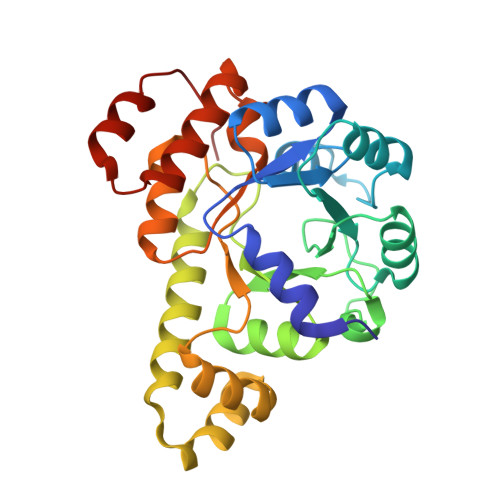
    Infectious diseases are a significant cause of death, and recent studies estimate that common bacterial infectious diseases were responsible for 13.6% of all global deaths in 2019. Among the most significant bacterial pathogens is Staphylococcus aureus, accounting for more than 1.1 million deaths worldwide in 2019. Vitamin biosynthesis has been proposed as a promising target for antibacterial therapy. Here, we investigated the biochemical, structural, and dynamic properties of the enzyme complex responsible for vitamin B6 (pyridoxal 5-phosphate, PLP) biosynthesis in S. aureus, which comprises enzymes SaPdx1 and SaPdx2. The crystal structure of the 24-mer complex of SaPdx1-SaPdx2 enzymes indicated that the S. aureus PLP synthase complex forms a highly dynamic assembly with transient interaction between the enzymes. Solution scattering data indicated that SaPdx2 typically binds to SaPdx1 at a substoichiometric ratio. We propose a structure-based view of the PLP synthesis mechanism initiated with the assembly of SaPLP synthase complex that proceeds in a highly dynamic interaction between Pdx1 and Pdx2. This interface interaction can be further explored as a potentially druggable site for the design of new antibiotics.


  • Organizational Affiliation

    São Carlos Institute of Physics, University of São Paulo, São Carlos, Brazil; Institute of Biochemistry and Molecular Biology, Laboratory for Structural Biology of Infection and Inflammation, University of Hamburg, Hamburg, Germany.


Macromolecules
Find similar proteins by:  (by identity cutoff)  |  3D Structure
Entity ID: 1
MoleculeChains Sequence LengthOrganismDetailsImage
Pyridoxal 5'-phosphate synthase subunit PdxS
A, B, C, D, E
A, B, C, D, E, F, G, H
274Staphylococcus aureusMutation(s): 0 
Gene Names: pdxS
UniProt
Find proteins for P60798 (Staphylococcus aureus (strain N315))
Explore P60798 
Go to UniProtKB:  P60798
Entity Groups  
Sequence Clusters30% Identity50% Identity70% Identity90% Identity95% Identity100% Identity
UniProt GroupP60798
Sequence Annotations
Expand
  • Reference Sequence
Small Molecules
Ligands 4 Unique
IDChains Name / Formula / InChI Key2D Diagram3D Interactions
PO4 (Subject of Investigation/LOI)
Query on PO4

Download Ideal Coordinates CCD File 
CA [auth F]
DA [auth F]
FA [auth G]
GA [auth G]
I [auth A]
CA [auth F],
DA [auth F],
FA [auth G],
GA [auth G],
I [auth A],
IA [auth H],
J [auth A],
JA [auth H],
N [auth B],
O [auth B],
R [auth C],
S [auth C],
U [auth D],
V [auth D],
X [auth E],
Y [auth E]
PHOSPHATE ION
O4 P
NBIIXXVUZAFLBC-UHFFFAOYSA-K
EDO
Query on EDO

Download Ideal Coordinates CCD File 
K [auth A],
P [auth B]
1,2-ETHANEDIOL
C2 H6 O2
LYCAIKOWRPUZTN-UHFFFAOYSA-N
CL
Query on CL

Download Ideal Coordinates CCD File 
AA [auth E]
EA [auth F]
KA [auth H]
L [auth A]
Q [auth B]
AA [auth E],
EA [auth F],
KA [auth H],
L [auth A],
Q [auth B],
T [auth C],
W [auth D],
Z [auth E]
CHLORIDE ION
Cl
VEXZGXHMUGYJMC-UHFFFAOYSA-M
MG
Query on MG

Download Ideal Coordinates CCD File 
BA [auth E],
HA [auth G],
M [auth A]
MAGNESIUM ION
Mg
JLVVSXFLKOJNIY-UHFFFAOYSA-N
Experimental Data & Validation

Experimental Data

  • Method: X-RAY DIFFRACTION
  • Resolution: 2.83 Å
  • R-Value Free: 0.260 
  • R-Value Work: 0.202 
  • R-Value Observed: 0.205 
  • Space Group: H 3 2
Unit Cell:
Length ( Å )Angle ( ˚ )
a = 192.419α = 90
b = 192.419β = 90
c = 448.198γ = 120
Software Package:
Software NamePurpose
PHENIXphasing
PHENIXrefinement
XDSdata reduction
SCALAdata scaling
Cootmodel building

Structure Validation

View Full Validation Report


Ligand Structure Quality Assessment 


Entry History & Funding Information

Deposition Data


Funding OrganizationLocationGrant Number
German Research Foundation (DFG)Germany390715994

Revision History  (Full details and data files)

  • Version 1.0: 2024-10-02
    Type: Initial release