8QNC | pdb_00008qnc

Crystal structure of ancestral L-galactono-1,4-lactone dehydrogenase: A113G variant


Experimental Data Snapshot

  • Method: X-RAY DIFFRACTION
  • Resolution: 1.70 Å
  • R-Value Free: 
    0.212 (Depositor), 0.222 (DCC) 
  • R-Value Work: 
    0.179 (Depositor), 0.191 (DCC) 
  • R-Value Observed: 
    0.180 (Depositor) 

Starting Model: in silico
View more details

wwPDB Validation 3D Report Full Report

Validation slider image for 8QNC

Ligand Structure Quality Assessment 


This is version 1.1 of the entry. See complete history

Literature

Structure, mechanism, and evolution of the last step in vitamin C biosynthesis.

Boverio, A.Jamil, N.Mannucci, B.Mascotti, M.L.Fraaije, M.W.Mattevi, A.

(2024) Nat Commun 15: 4158-4158

  • DOI: https://doi.org/10.1038/s41467-024-48410-1
  • Primary Citation Related Structures: 
    8QMY, 8QNB, 8QNC, 8QNR

  • PubMed Abstract: 

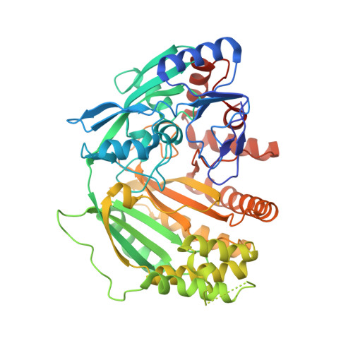
    Photosynthetic organisms, fungi, and animals comprise distinct pathways for vitamin C biosynthesis. Besides this diversity, the final biosynthetic step consistently involves an oxidation reaction carried out by the aldonolactone oxidoreductases. Here, we study the origin and evolution of the diversified activities and substrate preferences featured by these flavoenzymes using molecular phylogeny, kinetics, mutagenesis, and crystallographic experiments. We find clear evidence that they share a common ancestor. A flavin-interacting amino acid modulates the reactivity with the electron acceptors, including oxygen, and determines whether an enzyme functions as an oxidase or a dehydrogenase. We show that a few side chains in the catalytic cavity impart the reaction stereoselectivity. Ancestral sequence reconstruction outlines how these critical positions were affixed to specific amino acids along the evolution of the major eukaryotic clades. During Eukarya evolution, the aldonolactone oxidoreductases adapted to the varying metabolic demands while retaining their overarching vitamin C-generating function.


  • Organizational Affiliation
    • Molecular Enzymology group, University of Groningen, Nijenborgh 4, 9747AG, Groningen, The Netherlands.

Macromolecule Content 

  • Total Structure Weight: 58.32 kDa 
  • Atom Count: 4,201 
  • Modeled Residue Count: 486 
  • Deposited Residue Count: 511 
  • Unique protein chains: 1

Macromolecules

Find similar proteins by:|  3D Structure
Entity ID: 1
MoleculeChains  Sequence LengthOrganismDetailsImage
L-galactono-1,4-lactone dehydrogenase: A113G variant511synthetic constructMutation(s): 0 
EC: 1.3.2.3

Experimental Data & Validation

Experimental Data

  • Method: X-RAY DIFFRACTION
  • Resolution: 1.70 Å
  • R-Value Free:  0.212 (Depositor), 0.222 (DCC) 
  • R-Value Work:  0.179 (Depositor), 0.191 (DCC) 
  • R-Value Observed: 0.180 (Depositor) 
Space Group: P 1 21 1
Unit Cell:
Length ( Å )Angle ( ˚ )
a = 53.387α = 90
b = 69.657β = 98.29
c = 67.608γ = 90
Software Package:
Software NamePurpose
REFMACrefinement
Aimlessdata scaling

Structure Validation

View Full Validation Report


Ligand Structure Quality Assessment 


Entry History 

& Funding Information

Deposition Data


Funding OrganizationLocationGrant Number
Not fundedItaly--

Revision History  (Full details and data files)

  • Version 1.0: 2024-05-01
    Type: Initial release
  • Version 1.1: 2024-11-13
    Changes: Database references, Structure summary