8QN3 | pdb_00008qn3

OPR3 wildtype in complex with NADH4


Experimental Data Snapshot

  • Method: X-RAY DIFFRACTION
  • Resolution: 1.75 Å
  • R-Value Free: 
    0.225 (Depositor), 0.225 (DCC) 
  • R-Value Work: 
    0.200 (Depositor), 0.200 (DCC) 
  • R-Value Observed: 
    0.202 (Depositor) 

Starting Model: experimental
View more details

wwPDB Validation 3D Report Full Report

Validation slider image for 8QN3

Ligand Structure Quality Assessment 


This is version 1.3 of the entry. See complete history

Literature

Loop 6 and the beta-hairpin flap are structural hotspots that determine cofactor specificity in the FMN-dependent family of ene-reductases.

Kerschbaumer, B.Totaro, M.G.Friess, M.Breinbauer, R.Bijelic, A.Macheroux, P.

(2024) FEBS J 291: 1560-1574

  • DOI: https://doi.org/10.1111/febs.17055
  • Primary Citation Related Structures: 
    8QMX, 8QN3

  • PubMed Abstract: 

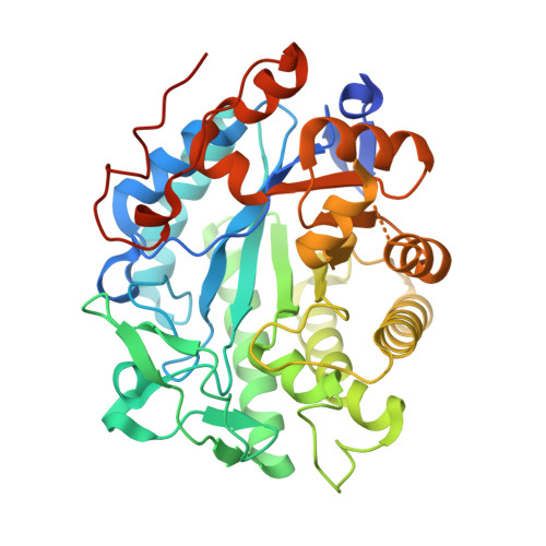
    Flavin mononucleotide (FMN)-dependent ene-reductases constitute a large family of oxidoreductases that catalyze the enantiospecific reduction of carbon-carbon double bonds. The reducing equivalents required for substrate reduction are obtained from reduced nicotinamide by hydride transfer. Most ene-reductases significantly prefer, or exclusively accept, either NADPH or NADH. Despite their usefulness in biocatalytic applications, the structural determinants for cofactor preference remain elusive. We employed the NADPH-preferring 12-oxophytodienoic acid reductase 3 from Solanum lycopersicum (SlOPR3) as a model enzyme of the ene-reductase family and applied computational and structural methods to investigate the binding specificity of the reducing coenzymes. Initial docking results indicated that the arginine triad R283, R343, and R366 residing on and close to a critical loop at the active site (loop 6) are the main contributors to NADPH binding. In contrast, NADH binds unfavorably in the opposite direction toward the β-hairpin flap within a largely hydrophobic region. Notably, the crystal structures of SlOPR3 in complex with either NADPH 4 or NADH 4 corroborated these different binding modes. Molecular dynamics simulations confirmed NADH binding near the β-hairpin flap and provided structural explanations for the low binding affinity of NADH to SlOPR3. We postulate that cofactor specificity is determined by the arginine triad/loop 6 and the residue(s) controlling access to a hydrophobic cleft formed by the β-hairpin flap. Thus, NADPH preference depends on a properly positioned arginine triad, whereas granting access to the hydrophobic cleft at the β-hairpin flap favors NADH binding.


  • Organizational Affiliation
    • Institute of Biochemistry, Graz University of Technology, Austria.

Macromolecule Content 

  • Total Structure Weight: 91.24 kDa 
  • Atom Count: 5,821 
  • Modeled Residue Count: 719 
  • Deposited Residue Count: 804 
  • Unique protein chains: 1

Macromolecules

Find similar proteins by:|  3D Structure
Entity ID: 1
MoleculeChains  Sequence LengthOrganismDetailsImage
12-oxophytodienoate reductase 3
A, B
402Solanum lycopersicumMutation(s): 0 
Gene Names: OPR3
EC: 1.3.1.42
UniProt
Find proteins for Q9FEW9 (Solanum lycopersicum)
Explore Q9FEW9 
Go to UniProtKB:  Q9FEW9
Entity Groups
Sequence Clusters30% Identity50% Identity70% Identity90% Identity95% Identity100% Identity
UniProt GroupQ9FEW9
Sequence Annotations
Expand
Reference Sequence

Small Molecules

Ligands 3 Unique
IDChains Name / Formula / InChI Key2D Diagram3D Interactions
WI6
(Subject of Investigation/LOI)

Query on WI6


Download:Ideal Coordinates CCD File
E [auth A],
H [auth B]
1,4,5,6-Tetrahydronicotinamide adenine dinucleotide
C21 H31 N7 O14 P2
GFDCQHGWOHYWLP-XQYAFYQJSA-N
FMN

Query on FMN


Download:Ideal Coordinates CCD File
C [auth A],
G [auth B]
FLAVIN MONONUCLEOTIDE
C17 H21 N4 O9 P
FVTCRASFADXXNN-SCRDCRAPSA-N
SO4

Query on SO4


Download:Ideal Coordinates CCD File
D [auth A],
F [auth A]
SULFATE ION
O4 S
QAOWNCQODCNURD-UHFFFAOYSA-L

Experimental Data & Validation

Experimental Data

  • Method: X-RAY DIFFRACTION
  • Resolution: 1.75 Å
  • R-Value Free:  0.225 (Depositor), 0.225 (DCC) 
  • R-Value Work:  0.200 (Depositor), 0.200 (DCC) 
  • R-Value Observed: 0.202 (Depositor) 
Space Group: P 1 21 1
Unit Cell:
Length ( Å )Angle ( ˚ )
a = 57.74α = 90
b = 89.46β = 107.401
c = 81.02γ = 90
Software Package:
Software NamePurpose
PHENIXrefinement
PHENIXrefinement
XDSdata reduction
XSCALEdata scaling
PHASERphasing

Structure Validation

View Full Validation Report


Ligand Structure Quality Assessment 


Entry History 

& Funding Information

Deposition Data


Funding OrganizationLocationGrant Number
Austrian Science FundAustria--

Revision History  (Full details and data files)

  • Version 1.0: 2024-01-17
    Type: Initial release
  • Version 1.1: 2024-02-07
    Changes: Database references
  • Version 1.2: 2024-04-10
    Changes: Database references
  • Version 1.3: 2025-02-05
    Changes: Data collection, Structure summary