8QJK | pdb_00008qjk

Structure of the cytoplasmic domain of csx23 from Vibrio cholera in complex with cyclic tetra-adenylate (cA4)


Experimental Data Snapshot

  • Method: X-RAY DIFFRACTION
  • Resolution: 1.76 Å
  • R-Value Free: 
    0.272 (Depositor), 0.270 (DCC) 
  • R-Value Work: 
    0.243 (Depositor), 0.248 (DCC) 

Starting Model: in silico
View more details

wwPDB Validation 3D Report Full Report

Validation slider image for 8QJK

This is version 1.1 of the entry. See complete history

Literature

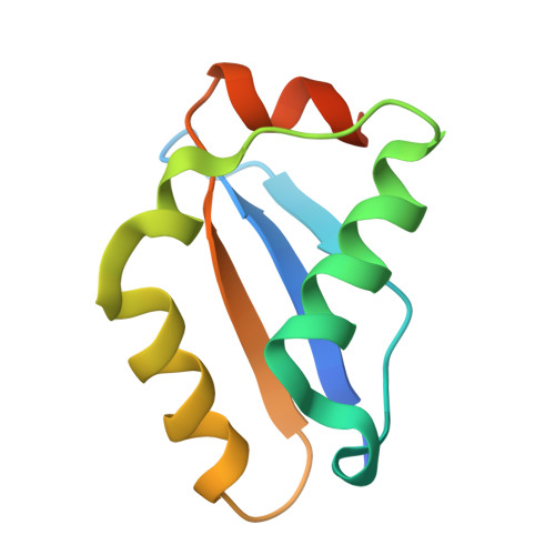
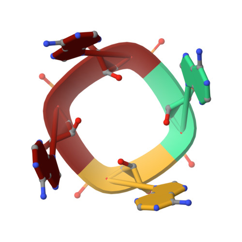
CRISPR antiphage defence mediated by the cyclic nucleotide-binding membrane protein Csx23.

Gruschow, S.McQuarrie, S.Ackermann, K.McMahon, S.Bode, B.E.Gloster, T.M.White, M.F.

(2024) Nucleic Acids Res 52: 2761-2775

  • DOI: https://doi.org/10.1093/nar/gkae167
  • Primary Citation Related Structures: 
    8QJK

  • PubMed Abstract: 

    CRISPR-Cas provides adaptive immunity in prokaryotes. Type III CRISPR systems detect invading RNA and activate the catalytic Cas10 subunit, which generates a range of nucleotide second messengers to signal infection. These molecules bind and activate a diverse range of effector proteins that provide immunity by degrading viral components and/or by disturbing key aspects of cellular metabolism to slow down viral replication. Here, we focus on the uncharacterised effector Csx23, which is widespread in Vibrio cholerae. Csx23 provides immunity against plasmids and phage when expressed in Escherichia coli along with its cognate type III CRISPR system. The Csx23 protein localises in the membrane using an N-terminal transmembrane α-helical domain and has a cytoplasmic C-terminal domain that binds cyclic tetra-adenylate (cA4), activating its defence function. Structural studies reveal a tetrameric structure with a novel fold that binds cA4 specifically. Using pulse EPR, we demonstrate that cA4 binding to the cytoplasmic domain of Csx23 results in a major perturbation of the transmembrane domain, consistent with the opening of a pore and/or disruption of membrane integrity. This work reveals a new class of cyclic nucleotide binding protein and provides key mechanistic detail on a membrane-associated CRISPR effector.


  • Organizational Affiliation
    • Biomedical Sciences Research Complex, School of Biology, University of St Andrews, St Andrews, Fife KY16 9ST, UK.

Macromolecule Content 

  • Total Structure Weight: 11.78 kDa 
  • Atom Count: 759 
  • Modeled Residue Count: 86 
  • Deposited Residue Count: 100 
  • Unique protein chains: 1
  • Unique nucleic acid chains: 1

Macromolecules


Find similar proteins by:|  3D Structure
Entity ID: 1
MoleculeChains  Sequence LengthOrganismDetailsImage
csx2396Vibrio choleraeMutation(s): 0 
UniProt
Find proteins for A0AAX7FLT5 (Vibrio cholerae)
Explore A0AAX7FLT5 
Go to UniProtKB:  A0AAX7FLT5
Entity Groups
Sequence Clusters30% Identity50% Identity70% Identity90% Identity95% Identity100% Identity
UniProt GroupA0AAX7FLT5
Sequence Annotations
Expand
Reference Sequence
Find similar nucleic acids by:  Sequence
Entity ID: 2
MoleculeChains LengthOrganismImage
Cyclic tetraadenosine monophosphate (cA4)B [auth C]4synthetic construct
Sequence Annotations
Expand
Reference Sequence

Biologically Interesting Molecules (External Reference) 

1 Unique

Experimental Data & Validation

Experimental Data

  • Method: X-RAY DIFFRACTION
  • Resolution: 1.76 Å
  • R-Value Free:  0.272 (Depositor), 0.270 (DCC) 
  • R-Value Work:  0.243 (Depositor), 0.248 (DCC) 
Space Group: I 4
Unit Cell:
Length ( Å )Angle ( ˚ )
a = 66.425α = 90
b = 66.425β = 90
c = 42.479γ = 90
Software Package:
Software NamePurpose
REFMACrefinement
autoPROCdata reduction
STARANISOdata scaling
PHASERphasing

Structure Validation

View Full Validation Report


Entry History 

& Funding Information

Deposition Data


Funding OrganizationLocationGrant Number
Biotechnology and Biological Sciences Research Council (BBSRC)United KingdomBB/T004789/1

Revision History  (Full details and data files)

  • Version 1.0: 2024-08-07
    Type: Initial release
  • Version 1.1: 2025-02-26
    Changes: Database references, Structure summary