A Fragment-Based Competitive 19 F LB-NMR Platform For Hotspot-Directed Ligand Profiling.
McCarthy, W.J., Thomas, S.E., Olaleye, T., Boland, J.A., Floto, R.A., Williams, G., Blundell, T.L., Coyne, A.G., Abell, C.(2024) Angew Chem Int Ed Engl 63: e202406846-e202406846
- PubMed: 38896426 Search on PubMed
- DOI: https://doi.org/10.1002/anie.202406846
- Primary Citation Related Structures:
8QID, 8QIX, 8QIY, 8QJ8 - PubMed Abstract:
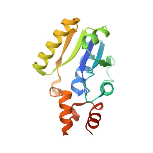
Ligand binding hotspots are regions of protein surfaces that form particularly favourable interactions with small molecule pharmacophores. Targeting interactions with these hotspots maximises the efficiency of ligand binding. Existing methods are capable of identifying hotspots but often lack assays to quantify ligand binding and direct elaboration at these sites. Herein, we describe a fragment-based competitive 19F Ligand Based-NMR (LB-NMR) screening platform that enables routine, quantitative ligand profiling focused at ligand-binding hotspots. As a proof of concept, the method was applied to 4'-phosphopantetheine adenylyltransferase (PPAT) from Mycobacterium abscessus (Mabs). X-ray crystallographic characterisation of the hits from a 960-member fragment screen identified three ligand-binding hotspots across the PPAT active site. From the fragment hits a collection of 19F reporter candidates were designed and synthesised. By rigorous prioritisation and use of optimisation workflows, a single 19F reporter molecule was generated for each hotspot. Profiling the binding of a set of structurally characterised ligands by competitive 19F LB-NMR with this suite of 19F reporters recapitulated the binding affinity and site ID assignments made by ITC and X-ray crystallography. This quantitative mapping of ligand binding events at hotspot level resolution establishes the utility of the fragment-based competitive 19F LB-NMR screening platform for hotspot-directed ligand profiling.
- University of Cambridge, Yusuf Hamid Department of Chemistry, UNITED KINGDOM.
Organizational Affiliation: