Capturing the blue-light activated state of the Phot-LOV1 domain from Chlamydomonas reinhardtii using time-resolved serial synchrotron crystallography.
Gotthard, G., Mous, S., Weinert, T., Maia, R.N.A., James, D., Dworkowski, F., Gashi, D., Furrer, A., Ozerov, D., Panepucci, E., Wang, M., Schertler, G.F.X., Heberle, J., Standfuss, J., Nogly, P.(2024) IUCrJ 11: 792-808
- PubMed: 39037420 Search on PubMed
- DOI: https://doi.org/10.1107/S2052252524005608
- Primary Citation Related Structures:
8QI8, 8QI9, 8QIA, 8QIB, 8QIF, 8QIG, 8QIH, 8QII, 8QIK, 8QIL, 8QIM, 8QIN, 8QIO, 8QIP, 8QIQ, 8QIR, 8QIS, 8QIT, 8QIU, 8QIV, 8QIW - PubMed Abstract:
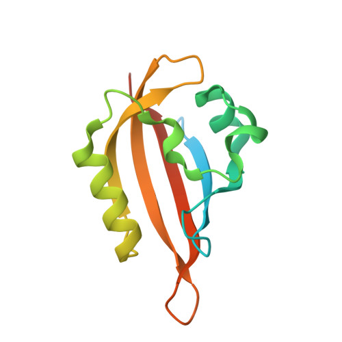
Light-oxygen-voltage (LOV) domains are small photosensory flavoprotein modules that allow the conversion of external stimuli (sunlight) into intracellular signals responsible for various cell behaviors (e.g. phototropism and chloroplast relocation). This ability relies on the light-induced formation of a covalent thioether adduct between a flavin chromophore and a reactive cysteine from the protein environment, which triggers a cascade of structural changes that result in the activation of a serine/threonine (Ser/Thr) kinase. Recent developments in time-resolved crystallography may allow the activation cascade of the LOV domain to be observed in real time, which has been elusive. In this study, we report a robust protocol for the production and stable delivery of microcrystals of the LOV domain of phototropin Phot-1 from Chlamydomonas reinhardtii (CrPhotLOV1) with a high-viscosity injector for time-resolved serial synchrotron crystallography (TR-SSX). The detailed process covers all aspects, from sample optimization to data collection, which may serve as a guide for soluble protein preparation for TR-SSX. In addition, we show that the crystals obtained preserve the photoreactivity using infrared spectroscopy. Furthermore, the results of the TR-SSX experiment provide high-resolution insights into structural alterations of CrPhotLOV1 from Δt = 2.5 ms up to Δt = 95 ms post-photoactivation, including resolving the geometry of the thioether adduct and the C-terminal region implicated in the signal transduction process.
- Laboratory of Biomolecular Research, Division of Biology and Chemistry, Paul Scherrer Institute, 5232 Villigen PSI, Switzerland.
Organizational Affiliation: