8QCH | pdb_00008qch

Human Adenosine deaminase-like protein in complex with compound AT8001


Experimental Data Snapshot

  • Method: X-RAY DIFFRACTION
  • Resolution: 2.44 Å
  • R-Value Free: 
    0.243 (Depositor), 0.233 (DCC) 
  • R-Value Work: 
    0.214 (Depositor), 0.202 (DCC) 
  • R-Value Observed: 
    0.216 (Depositor) 

Starting Model: experimental
View more details

wwPDB Validation 3D Report Full Report

Validation slider image for 8QCH

Ligand Structure Quality Assessment 


This is version 1.1 of the entry. See complete history

Literature

The activation cascade of the broad-spectrum antiviral bemnifosbuvir characterized at atomic resolution.

Chazot, A.Zimberger, C.Feracci, M.Moussa, A.Good, S.Sommadossi, J.P.Alvarez, K.Ferron, F.Canard, B.

(2024) PLoS Biol 22: e3002743-e3002743

  • DOI: https://doi.org/10.1371/journal.pbio.3002743
  • Primary Citation Related Structures: 
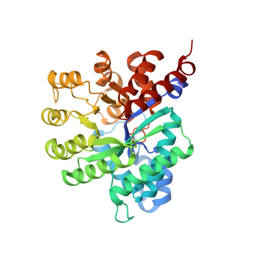
    8PIE, 8PTS, 8PWK, 8QCH

  • PubMed Abstract: 

    Bemnifosbuvir (AT-527) and AT-752 are guanosine analogues currently in clinical trials against several RNA viruses. Here, we show that these drugs require a minimal set of 5 cellular enzymes for activation to their common 5'-triphosphate AT-9010, with an obligate order of reactions. AT-9010 selectively inhibits essential viral enzymes, accounting for antiviral potency. Functional and structural data at atomic resolution decipher N6-purine deamination compatible with its metabolic activation. Crystal structures of human histidine triad nucleotide binding protein 1, adenosine deaminase-like protein 1, guanylate kinase 1, and nucleoside diphosphate kinase at 2.09, 2.44, 1.76, and 1.9 Å resolution, respectively, with cognate precursors of AT-9010 illuminate the activation pathway from the orally available bemnifosbuvir to AT-9010, pointing to key drug-protein contacts along the activation pathway. Our work provides a framework to integrate the design of antiviral nucleotide analogues, confronting requirements and constraints associated with activation enzymes along the 5'-triphosphate assembly line.


  • Organizational Affiliation
    • Aix Marseille Université, CNRS, AFMB, UMR 7257, Marseille, France.

Macromolecule Content 

  • Total Structure Weight: 328.2 kDa 
  • Atom Count: 23,207 
  • Modeled Residue Count: 2,765 
  • Deposited Residue Count: 2,840 
  • Unique protein chains: 1

Macromolecules

Find similar proteins by:|  3D Structure
Entity ID: 1
MoleculeChains  Sequence LengthOrganismDetailsImage
Adenosine deaminase-like protein
A, B, C, D, E
A, B, C, D, E, F, G, H
355Homo sapiensMutation(s): 0 
Gene Names: ADALADAL1
EC: 3.5.4
UniProt & NIH Common Fund Data Resources
Find proteins for Q6DHV7 (Homo sapiens)
Explore Q6DHV7 
Go to UniProtKB:  Q6DHV7
PHAROS:  Q6DHV7
GTEx:  ENSG00000168803 
Entity Groups
Sequence Clusters30% Identity50% Identity70% Identity90% Identity95% Identity100% Identity
UniProt GroupQ6DHV7
Sequence Annotations
Expand
Reference Sequence

Small Molecules

Ligands 5 Unique
IDChains Name / Formula / InChI Key2D Diagram3D Interactions
EIF
(Subject of Investigation/LOI)

Query on EIF


Download:Ideal Coordinates CCD File
CA [auth E]
GA [auth F]
I [auth A]
N [auth B]
NA [auth G]
CA [auth E],
GA [auth F],
I [auth A],
N [auth B],
NA [auth G],
QA [auth H],
R [auth C],
X [auth D]
[(2~{R},3~{R},4~{R},5~{R})-5-(2-azanyl-6-oxidanylidene-1~{H}-purin-9-yl)-4-fluoranyl-4-methyl-3-oxidanyl-oxolan-2-yl]methyl dihydrogen phosphate
C11 H15 F N5 O7 P
BOEZKCHYGLAVGV-GITKWUPZSA-N
PEG

Query on PEG


Download:Ideal Coordinates CCD File
BA [auth E],
MA [auth G],
V [auth D],
W [auth D]
DI(HYDROXYETHYL)ETHER
C4 H10 O3
MTHSVFCYNBDYFN-UHFFFAOYSA-N
SO4

Query on SO4


Download:Ideal Coordinates CCD File
AA [auth D]
EA [auth E]
FA [auth E]
IA [auth F]
JA [auth F]
AA [auth D],
EA [auth E],
FA [auth E],
IA [auth F],
JA [auth F],
K [auth A],
KA [auth F],
L [auth A],
LA [auth F],
M [auth A],
P [auth B],
PA [auth G],
Q [auth B],
SA [auth H],
T [auth C],
U [auth C],
Z [auth D]
SULFATE ION
O4 S
QAOWNCQODCNURD-UHFFFAOYSA-L
ZN
(Subject of Investigation/LOI)

Query on ZN


Download:Ideal Coordinates CCD File
DA [auth E]
HA [auth F]
J [auth A]
O [auth B]
OA [auth G]
DA [auth E],
HA [auth F],
J [auth A],
O [auth B],
OA [auth G],
RA [auth H],
S [auth C],
Y [auth D]
ZINC ION
Zn
PTFCDOFLOPIGGS-UHFFFAOYSA-N
NA

Query on NA


Download:Ideal Coordinates CCD File
TA [auth H]SODIUM ION
Na
FKNQFGJONOIPTF-UHFFFAOYSA-N

Experimental Data & Validation

Experimental Data

  • Method: X-RAY DIFFRACTION
  • Resolution: 2.44 Å
  • R-Value Free:  0.243 (Depositor), 0.233 (DCC) 
  • R-Value Work:  0.214 (Depositor), 0.202 (DCC) 
  • R-Value Observed: 0.216 (Depositor) 
Space Group: P 21 21 2
Unit Cell:
Length ( Å )Angle ( ˚ )
a = 142.757α = 90
b = 142.983β = 90
c = 156.399γ = 90
Software Package:
Software NamePurpose
BUSTERrefinement
autoPROCdata reduction
Aimlessdata scaling
MoRDaphasing

Structure Validation

View Full Validation Report


Ligand Structure Quality Assessment 


Entry History 

& Funding Information

Deposition Data


Funding OrganizationLocationGrant Number
Agence Nationale de la Recherche (ANR)FranceANR-10-INBS-0005
Fondation pour la Recherche Medicale (FRM)FranceSoutien aux equipes

Revision History  (Full details and data files)

  • Version 1.0: 2024-07-31
    Type: Initial release
  • Version 1.1: 2024-09-04
    Changes: Database references