8Q8H | pdb_00008q8h

Crystal Structure of Apo beta-D-GalNAcase from Niabella aurantiaca (Structure 2)


Experimental Data Snapshot

  • Method: X-RAY DIFFRACTION
  • Resolution: 2.50 Å
  • R-Value Free: 
    0.232 (Depositor), 0.198 (DCC) 
  • R-Value Work: 
    0.188 (Depositor), 0.196 (DCC) 
  • R-Value Observed: 
    0.190 (Depositor) 

Starting Model: in silico
View more details

wwPDB Validation 3D Report Full Report

Validation slider image for 8Q8H

Ligand Structure Quality Assessment 


This is version 1.0 of the entry. See complete history

Literature

Characterization and structural study of a novel beta-N-acetylgalactosaminidase from Niabella aurantiaca.

Moreno Prieto, E.S.Fjermedal, S.Siebenhaar, S.Vuillemin, M.Holck, J.Vincentelli, R.Gippert, G.P.Wilkens, C.Morth, J.P.Henrissat, B.

(2024) FEBS J 291: 1439-1456

  • DOI: https://doi.org/10.1111/febs.17042
  • Primary Citation Related Structures: 
    8P4L, 8Q8H

  • PubMed Abstract: 

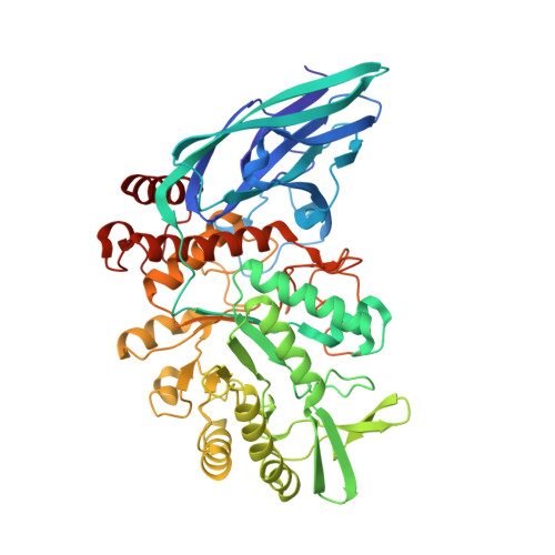
    We report here the identification, characterization and three-dimensional (3D) structure determination of NaNga, a newly identified β-N-acetylgalactosaminidase from the Gram-negative soil bacterium Niabella aurantiaca DSM 17617. When recombinantly expressed in Escherichia coli, the enzyme selectively cleaved 4-nitrophenyl-N-acetyl-β-d-galactosamine (pNP-β-d-GalpNAc). The X-ray crystal structure of the protein was refined to 2.5 Å and consists of an N-terminal β-sandwich domain and a (β/α) 8 barrel catalytic domain. Despite a mere 22% sequence identity, the 3D structure of NaNga is similar to those previously determined for family GH123 members, suggesting it also employs the same substrate-assisted catalytic mechanism. Inhibition by N-acetyl-galactosamine thiazoline (GalNAc-thiazoline) supports the suggested mechanism. A phylogenetic analysis of its proximal sequence space shows significant clustering of unknown sequences around NaNga with sufficient divergence with previously identified GH123 members to subdivide this family into distinct subfamilies. Although the actual biological substrate of our enzyme remains unknown, examination of the active site pocket suggests that it may be a β-N-acetylgalactosaminide substituted by a monosaccharide at O-3. Analysis of the genomic context suggests, in turn, that this substituted β-N-acetylgalactosaminide may be appended to a d-arabinan from an environmental Actinomycete.


  • Organizational Affiliation
    • Department of Biotechnology and Biomedicine, Technical University of Denmark, Kgs. Lyngby, Denmark.

Macromolecule Content 

  • Total Structure Weight: 252.91 kDa 
  • Atom Count: 18,026 
  • Modeled Residue Count: 2,144 
  • Deposited Residue Count: 2,144 
  • Unique protein chains: 1

Macromolecules

Find similar proteins by:|  3D Structure
Entity ID: 1
MoleculeChains  Sequence LengthOrganismDetailsImage
beta-D-GalNAcase from Niabella aurantiaca DSM 17617
A, B, C, D
536Niabella aurantiaca DSM 17617Mutation(s): 0 
Gene Names: NZ_KB893637.1
EC: 3.2.1.53
UniProt
Find proteins for A0AAT8XUD7 (Niabella aurantiaca DSM 17617)
Explore A0AAT8XUD7 
Go to UniProtKB:  A0AAT8XUD7
Entity Groups
Sequence Clusters30% Identity50% Identity70% Identity90% Identity95% Identity100% Identity
UniProt GroupA0AAT8XUD7
Sequence Annotations
Expand
Reference Sequence

Small Molecules

Ligands 2 Unique
IDChains Name / Formula / InChI Key2D Diagram3D Interactions
SO4
(Subject of Investigation/LOI)

Query on SO4


Download:Ideal Coordinates CCD File
E [auth A]
F [auth A]
G [auth A]
I [auth B]
J [auth B]
E [auth A],
F [auth A],
G [auth A],
I [auth B],
J [auth B],
L [auth C],
M [auth C],
N [auth C],
O [auth C],
Q [auth D]
SULFATE ION
O4 S
QAOWNCQODCNURD-UHFFFAOYSA-L
NA

Query on NA


Download:Ideal Coordinates CCD File
H [auth A],
K [auth B],
P [auth C],
R [auth D]
SODIUM ION
Na
FKNQFGJONOIPTF-UHFFFAOYSA-N

Experimental Data & Validation

Experimental Data

  • Method: X-RAY DIFFRACTION
  • Resolution: 2.50 Å
  • R-Value Free:  0.232 (Depositor), 0.198 (DCC) 
  • R-Value Work:  0.188 (Depositor), 0.196 (DCC) 
  • R-Value Observed: 0.190 (Depositor) 
Space Group: P 21 21 2
Unit Cell:
Length ( Å )Angle ( ˚ )
a = 173.64α = 90
b = 195.57β = 90
c = 82.04γ = 90
Software Package:
Software NamePurpose
PHENIXrefinement
XDSdata reduction
XSCALEdata scaling
PHASERphasing

Structure Validation

View Full Validation Report


Ligand Structure Quality Assessment 


Entry History 

& Funding Information

Deposition Data


Funding OrganizationLocationGrant Number
Novo Nordisk FoundationDenmarkNNF20SA0067193

Revision History  (Full details and data files)

  • Version 1.0: 2024-05-15
    Type: Initial release