8Q5X

MgADP-bound Fe protein of the molybdenum nitrogenase from Methanococcus maripaludis


Experimental Data Snapshot

  • Method: X-RAY DIFFRACTION
  • Resolution: 1.70 Å
  • R-Value Free: 0.186 
  • R-Value Work: 0.160 
  • R-Value Observed: 0.162 

Starting Model: experimental
View more details

wwPDB Validation   3D Report Full Report


Ligand Structure Quality Assessment 


This is version 1.1 of the entry. See complete history


Literature

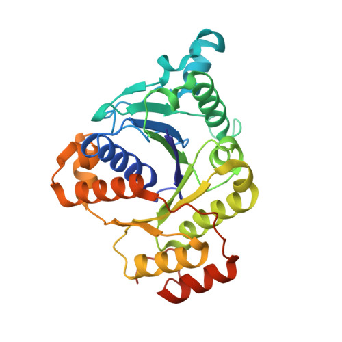
Structural comparison of (hyper-)thermophilic nitrogenase reductases from three marine Methanococcales.

Maslac, N.Cadoux, C.Bolte, P.Murken, F.Gu, W.Milton, R.D.Wagner, T.

(2024) FEBS J 291: 3454-3480

  • DOI: https://doi.org/10.1111/febs.17148
  • Primary Citation of Related Structures:  
    8Q50, 8Q5T, 8Q5V, 8Q5W, 8Q5X

  • PubMed Abstract: 

    The nitrogenase reductase NifH catalyses ATP-dependent electron delivery to the Mo-nitrogenase, a reaction central to biological dinitrogen (N 2 ) fixation. While NifHs have been extensively studied in bacteria, structural information about their archaeal counterparts is limited. Archaeal NifHs are considered more ancient, particularly those from Methanococcales, a group of marine hydrogenotrophic methanogens, which includes diazotrophs growing at temperatures near 92 °C. Here, we structurally and biochemically analyse NifHs from three Methanococcales, offering the X-ray crystal structures from meso-, thermo-, and hyperthermophilic methanogens. While NifH from Methanococcus maripaludis (37 °C) was obtained through heterologous recombinant expression, the proteins from Methanothermococcus thermolithotrophicus (65 °C) and Methanocaldococcus infernus (85 °C) were natively purified from the diazotrophic archaea. The structures from M. thermolithotrophicus crystallised as isolated exhibit high flexibility. In contrast, the complexes of NifH with MgADP obtained from the three methanogens are superposable, more rigid, and present remarkable structural conservation with their homologues. They retain key structural features of P-loop NTPases and share similar electrostatic profiles with the counterpart from the bacterial model organism Azotobacter vinelandii. In comparison to the NifH from the phylogenetically distant Methanosarcina acetivorans, these reductases do not cross-react significantly with Mo-nitrogenase from A. vinelandii. However, they associate with bacterial nitrogenase when ADP· AlF 4 - $$ {\mathrm{AlF}}_4^{-} $$ is added to mimic a transient reactive state. Accordingly, detailed surface analyses suggest that subtle substitutions would affect optimal binding during the catalytic cycle between the NifH from Methanococcales and the bacterial nitrogenase, implying differences in the N 2 -machinery from these ancient archaea.


  • Organizational Affiliation

    Microbial Metabolism Research Group, Max Planck Institute for Marine Microbiology, Bremen, Germany.


Macromolecules
Find similar proteins by:  (by identity cutoff)  |  3D Structure
Entity ID: 1
MoleculeChains Sequence LengthOrganismDetailsImage
Nitrogenase iron protein
A, B, C, D
288Methanococcus maripaludis S2Mutation(s): 0 
Gene Names: nifH
UniProt
Find proteins for P0CW57 (Methanococcus maripaludis (strain DSM 14266 / JCM 13030 / NBRC 101832 / S2 / LL))
Explore P0CW57 
Go to UniProtKB:  P0CW57
Entity Groups  
Sequence Clusters30% Identity50% Identity70% Identity90% Identity95% Identity100% Identity
UniProt GroupP0CW57
Sequence Annotations
Expand
  • Reference Sequence
Small Molecules
Ligands 8 Unique
IDChains Name / Formula / InChI Key2D Diagram3D Interactions
ADP (Subject of Investigation/LOI)
Query on ADP

Download Ideal Coordinates CCD File 
DA [auth D],
E [auth A],
N [auth B],
U [auth C]
ADENOSINE-5'-DIPHOSPHATE
C10 H15 N5 O10 P2
XTWYTFMLZFPYCI-KQYNXXCUSA-N
SF4 (Subject of Investigation/LOI)
Query on SF4

Download Ideal Coordinates CCD File 
G [auth A],
P [auth B]
IRON/SULFUR CLUSTER
Fe4 S4
LJBDFODJNLIPKO-UHFFFAOYSA-N
PEG
Query on PEG

Download Ideal Coordinates CCD File 
AA [auth C]DI(HYDROXYETHYL)ETHER
C4 H10 O3
MTHSVFCYNBDYFN-UHFFFAOYSA-N
GOL
Query on GOL

Download Ideal Coordinates CCD File 
GA [auth D],
Q [auth B],
Z [auth C]
GLYCEROL
C3 H8 O3
PEDCQBHIVMGVHV-UHFFFAOYSA-N
EDO
Query on EDO

Download Ideal Coordinates CCD File 
CA [auth D],
FA [auth D],
K [auth A],
Y [auth C]
1,2-ETHANEDIOL
C2 H6 O2
LYCAIKOWRPUZTN-UHFFFAOYSA-N
ACT
Query on ACT

Download Ideal Coordinates CCD File 
H [auth A]
HA [auth D]
I [auth A]
J [auth A]
R [auth B]
H [auth A],
HA [auth D],
I [auth A],
J [auth A],
R [auth B],
S [auth B],
W [auth C],
X [auth C]
ACETATE ION
C2 H3 O2
QTBSBXVTEAMEQO-UHFFFAOYSA-M
CA
Query on CA

Download Ideal Coordinates CCD File 
BA [auth D],
L [auth B],
M [auth B],
T [auth C]
CALCIUM ION
Ca
BHPQYMZQTOCNFJ-UHFFFAOYSA-N
MG (Subject of Investigation/LOI)
Query on MG

Download Ideal Coordinates CCD File 
EA [auth D],
F [auth A],
O [auth B],
V [auth C]
MAGNESIUM ION
Mg
JLVVSXFLKOJNIY-UHFFFAOYSA-N
Experimental Data & Validation

Experimental Data

  • Method: X-RAY DIFFRACTION
  • Resolution: 1.70 Å
  • R-Value Free: 0.186 
  • R-Value Work: 0.160 
  • R-Value Observed: 0.162 
  • Space Group: P 1 21 1
Unit Cell:
Length ( Å )Angle ( ˚ )
a = 68.546α = 90
b = 111.094β = 90.01
c = 78.82γ = 90
Software Package:
Software NamePurpose
PHENIXrefinement
autoPROCdata reduction
autoPROCdata scaling
PHASERphasing

Structure Validation

View Full Validation Report


Ligand Structure Quality Assessment 


Entry History & Funding Information

Deposition Data


Funding OrganizationLocationGrant Number
Max Planck SocietyGermany--
Swiss National Science FoundationSwitzerlandNCCR Catalysis 180544

Revision History  (Full details and data files)

  • Version 1.0: 2024-05-15
    Type: Initial release
  • Version 1.1: 2024-08-14
    Changes: Database references