8Q5U

Endoglycosidase S2 in complex with IgG1 Fc


Experimental Data Snapshot

  • Method: X-RAY DIFFRACTION
  • Resolution: 3.00 Å
  • R-Value Free: 0.252 
  • R-Value Work: 0.216 

Starting Models: experimental
View more details

wwPDB Validation   3D Report Full Report


This is version 1.1 of the entry. See complete history


Literature

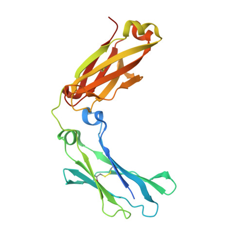
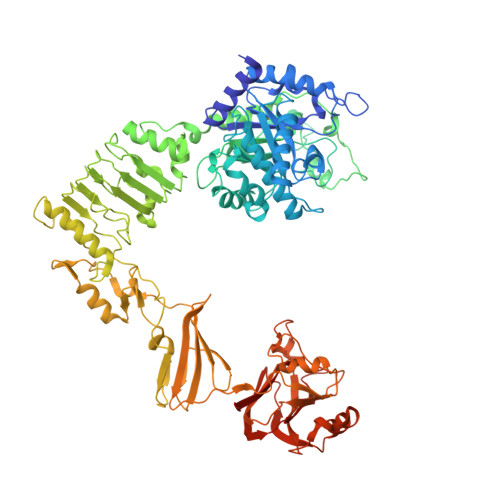
The IgG-specific endoglycosidases EndoS and EndoS2 are distinguished by conformation and antibody recognition.

Sudol, A.S.L.Crispin, M.Tews, I.

(2024) J Biol Chem 300: 107245-107245

  • DOI: https://doi.org/10.1016/j.jbc.2024.107245
  • Primary Citation of Related Structures:  
    8Q5U

  • PubMed Abstract: 

    The IgG-specific endoglycosidases EndoS and EndoS2 from Streptococcus pyogenes can remove conserved N-linked glycans present on the Fc region of host antibodies to inhibit Fc-mediated effector functions. These enzymes are therefore being investigated as therapeutics for suppressing unwanted immune activation, and have additional application as tools for antibody glycan remodeling. EndoS and EndoS2 differ in Fc glycan substrate specificity due to structural differences within their catalytic glycosyl hydrolase domains. However, a chimeric EndoS enzyme with a substituted glycosyl hydrolase from EndoS2 loses catalytic activity, despite high structural homology between the two enzymes, indicating either mechanistic divergence of EndoS and EndoS2, or improperly-formed domain interfaces in the chimeric enzyme. Here, we present the crystal structure of the EndoS2-IgG1 Fc complex determined to 3.0 Å resolution. Comparison of complexed and unliganded EndoS2 reveals relative reorientation of the glycosyl hydrolase, leucine-rich repeat and hybrid immunoglobulin domains. The conformation of the complexed EndoS2 enzyme is also different when compared to the earlier EndoS-IgG1 Fc complex, and results in distinct contact surfaces between the two enzymes and their Fc substrate. These findings indicate mechanistic divergence of EndoS2 and EndoS. It will be important to consider these differences in the design of IgG-specific enzymes, developed to enable customizable antibody glycosylation.


  • Organizational Affiliation

    School of Biological Sciences, University of Southampton, Southampton, UK.


Macromolecules
Find similar proteins by:  (by identity cutoff)  |  3D Structure
Entity ID: 1
MoleculeChains Sequence LengthOrganismDetailsImage
Uncharacterized protein DKFZp686C11235A,
C,
F [auth B]
227Homo sapiensMutation(s): 2 
Gene Names: DKFZp686C11235
UniProt
Find proteins for P0DOX5 (Homo sapiens)
Explore P0DOX5 
Go to UniProtKB:  P0DOX5
Entity Groups  
Sequence Clusters30% Identity50% Identity70% Identity90% Identity95% Identity100% Identity
UniProt GroupP0DOX5
Glycosylation
Glycosylation Sites: 1
Sequence Annotations
Expand
  • Reference Sequence
Find similar proteins by:  (by identity cutoff)  |  3D Structure
Entity ID: 2
MoleculeChains Sequence LengthOrganismDetailsImage
Endo-beta-N-acetylglucosaminidaseB [auth D],
D [auth E],
E [auth F]
816Streptococcus pyogenesMutation(s): 2 
Gene Names: endoS
UniProt
Find proteins for T1WGN1 (Streptococcus pyogenes serotype M49)
Explore T1WGN1 
Go to UniProtKB:  T1WGN1
Entity Groups  
Sequence Clusters30% Identity50% Identity70% Identity90% Identity95% Identity100% Identity
UniProt GroupT1WGN1
Sequence Annotations
Expand
  • Reference Sequence
Oligosaccharides

Help

Entity ID: 3
MoleculeChains Length2D Diagram Glycosylation3D Interactions
2-acetamido-2-deoxy-beta-D-glucopyranose-(1-2)-alpha-D-mannopyranose-(1-3)-[2-acetamido-2-deoxy-beta-D-glucopyranose-(1-2)-alpha-D-mannopyranose-(1-6)]beta-D-mannopyranose-(1-4)-2-acetamido-2-deoxy-beta-D-glucopyranose-(1-4)-[alpha-L-fucopyranose-(1-6)]2-acetamido-2-deoxy-beta-D-glucopyranose
G
8N-Glycosylation
Glycosylation Resources
GlyTouCan:  G80858MF
GlyCosmos:  G80858MF
GlyGen:  G80858MF
Entity ID: 4
MoleculeChains Length2D Diagram Glycosylation3D Interactions
2-acetamido-2-deoxy-beta-D-glucopyranose-(1-2)-alpha-D-mannopyranose-(1-6)-[alpha-D-mannopyranose-(1-3)]beta-D-mannopyranose-(1-4)-2-acetamido-2-deoxy-beta-D-glucopyranose-(1-4)-[alpha-L-fucopyranose-(1-6)]2-acetamido-2-deoxy-beta-D-glucopyranose
H
7N-Glycosylation
Glycosylation Resources
GlyTouCan:  G45889JQ
GlyCosmos:  G45889JQ
GlyGen:  G45889JQ
Small Molecules
Ligands 3 Unique
IDChains Name / Formula / InChI Key2D Diagram3D Interactions
PGE
Query on PGE

Download Ideal Coordinates CCD File 
J [auth D],
K [auth D],
L [auth D],
P [auth F],
Q [auth F]
TRIETHYLENE GLYCOL
C6 H14 O4
ZIBGPFATKBEMQZ-UHFFFAOYSA-N
GOL
Query on GOL

Download Ideal Coordinates CCD File 
I [auth A],
M [auth D],
S [auth B],
T [auth B]
GLYCEROL
C3 H8 O3
PEDCQBHIVMGVHV-UHFFFAOYSA-N
CA
Query on CA

Download Ideal Coordinates CCD File 
N [auth D],
O [auth E],
R [auth F]
CALCIUM ION
Ca
BHPQYMZQTOCNFJ-UHFFFAOYSA-N
Experimental Data & Validation

Experimental Data

  • Method: X-RAY DIFFRACTION
  • Resolution: 3.00 Å
  • R-Value Free: 0.252 
  • R-Value Work: 0.216 
  • Space Group: P 43 21 2
Unit Cell:
Length ( Å )Angle ( ˚ )
a = 228.775α = 90
b = 228.775β = 90
c = 161.629γ = 90
Software Package:
Software NamePurpose
REFMACrefinement
DIALSdata reduction
DIALSdata scaling
MOLREPphasing

Structure Validation

View Full Validation Report


Entry History & Funding Information

Deposition Data


Funding OrganizationLocationGrant Number
University of SouthamptonUnited Kingdom--

Revision History  (Full details and data files)

  • Version 1.0: 2024-04-17
    Type: Initial release
  • Version 1.1: 2024-05-08
    Changes: Database references