


Crystal structure of truncated human Microtubule-associated proteins 1A/1B light chain 3B in apo form
Kumar, A., Knapp, S.To be published.
Experimental Data Snapshot
Starting Model: experimental
View more details
wwPDB Validation 3D Report Full Report

Entity ID: 1 | |||||
|---|---|---|---|---|---|
| Molecule | Chains | Sequence Length | Organism | Details | Image |
| Microtubule-associated proteins 1A/1B light chain 3B | 126 | Homo sapiens | Mutation(s): 0 Gene Names: MAP1LC3B | 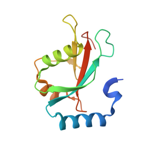 | |
UniProt & NIH Common Fund Data Resources | |||||
PHAROS: Q9GZQ8 GTEx: ENSG00000140941 | |||||
Entity Groups | |||||
| Sequence Clusters | 30% Identity50% Identity70% Identity90% Identity95% Identity100% Identity | ||||
| UniProt Group | Q9GZQ8 | ||||
Sequence AnnotationsExpand | |||||
Reference Sequence | |||||
| Ligands 2 Unique | |||||
|---|---|---|---|---|---|
| ID | Chains | Name / Formula / InChI Key | 2D Diagram | 3D Interactions | |
| EDO Download:Ideal Coordinates CCD File | B [auth A] C [auth A] D [auth A] E [auth A] F [auth A] | 1,2-ETHANEDIOL C2 H6 O2 LYCAIKOWRPUZTN-UHFFFAOYSA-N |  | ||
| CL Download:Ideal Coordinates CCD File | K [auth A] | CHLORIDE ION Cl VEXZGXHMUGYJMC-UHFFFAOYSA-M |  | ||
| Length ( Å ) | Angle ( ˚ ) |
|---|---|
| a = 60.255 | α = 90 |
| b = 60.255 | β = 90 |
| c = 34.927 | γ = 90 |
| Software Name | Purpose |
|---|---|
| PHENIX | refinement |
| XDS | data reduction |
| PHENIX | phasing |
| PHENIX | refinement |
| XDS | data scaling |
| Funding Organization | Location | Grant Number |
|---|---|---|
| European Union (EU) | European Union | 875510 |


