8Q3W | pdb_00008q3w

ATP-bound IstB in complex to duplex DNA


Experimental Data Snapshot

  • Method: ELECTRON MICROSCOPY
  • Resolution: 3.18 Å
  • Aggregation State: PARTICLE 
  • Reconstruction Method: SINGLE PARTICLE 

wwPDB Validation 3D Report Full Report

Validation slider image for 8Q3W

This is version 1.0 of the entry. See complete history

Literature

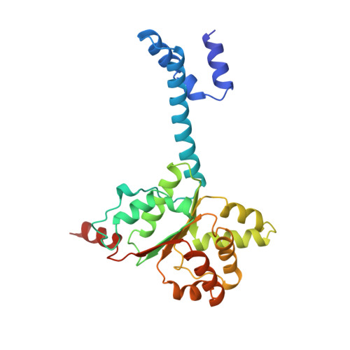
Molecular basis for transposase activation by a dedicated AAA+ ATPase.

de la Gandara, A.Spinola-Amilibia, M.Araujo-Bazan, L.Nunez-Ramirez, R.Berger, J.M.Arias-Palomo, E.

(2024) Nature 630: 1003-1011

  • DOI: https://doi.org/10.1038/s41586-024-07550-6
  • Primary Citation Related Structures: 
    8Q3W, 8Q4D

  • PubMed Abstract: 

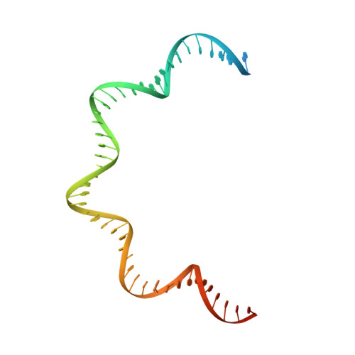
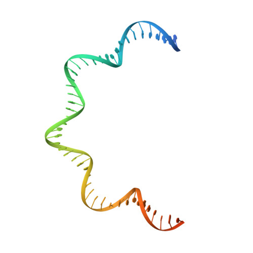
    Transposases drive chromosomal rearrangements and the dissemination of drug-resistance genes and toxins 1-3 . Although some transposases act alone, many rely on dedicated AAA+ ATPase subunits that regulate site selectivity and catalytic function through poorly understood mechanisms. Using IS21 as a model transposase system, we show how an ATPase regulator uses nucleotide-controlled assembly and DNA deformation to enable structure-based site selectivity, transposase recruitment, and activation and integration. Solution and cryogenic electron microscopy studies show that the IstB ATPase self-assembles into an autoinhibited pentamer of dimers that tightly curves target DNA into a half-coil. Two of these decamers dimerize, which stabilizes the target nucleic acid into a kinked S-shaped configuration that engages the IstA transposase at the interface between the two IstB oligomers to form an approximately 1 MDa transpososome complex. Specific interactions stimulate regulator ATPase activity and trigger a large conformational change on the transposase that positions the catalytic site to perform DNA strand transfer. These studies help explain how AAA+ ATPase regulators-which are used by classical transposition systems such as Tn7, Mu and CRISPR-associated elements-can remodel their substrate DNA and cognate transposases to promote function.


  • Organizational Affiliation
    • Centro de Investigaciones Biológicas Margarita Salas, CSIC, Madrid, Spain.

Macromolecule Content 

  • Total Structure Weight: 338.32 kDa 
  • Atom Count: 22,498 
  • Modeled Residue Count: 2,556 
  • Deposited Residue Count: 2,660 
  • Unique protein chains: 1
  • Unique nucleic acid chains: 2

Macromolecules


Find similar proteins by:|  3D Structure
Entity ID: 1
MoleculeChains  Sequence LengthOrganismDetailsImage
Insertion sequence IS5376 putative ATP-binding protein
A, B, C, D, E
A, B, C, D, E, F, G, H, I, J
254Geobacillus stearothermophilusMutation(s): 0 
UniProt
Find proteins for Q45619 (Geobacillus stearothermophilus)
Explore Q45619 
Go to UniProtKB:  Q45619
Entity Groups
Sequence Clusters30% Identity50% Identity70% Identity90% Identity95% Identity100% Identity
UniProt GroupQ45619
Sequence Annotations
Expand
Reference Sequence
Find similar nucleic acids by:  (by identity cutoff) 
Entity ID: 2
MoleculeChains LengthOrganismImage
DNA (48-MER) Target DNA FW60Geobacillus stearothermophilus
Sequence Annotations
Expand
Reference Sequence
Find similar nucleic acids by:  (by identity cutoff) 
Entity ID: 3
MoleculeChains LengthOrganismImage
DNA (48-MER) Traget DNA Rv60Geobacillus stearothermophilus
Sequence Annotations
Expand
Reference Sequence

Small Molecules

Ligands 2 Unique
IDChains Name / Formula / InChI Key2D Diagram3D Interactions
ATP
(Subject of Investigation/LOI)

Query on ATP


Download:Ideal Coordinates CCD File
BA [auth H]
DA [auth I]
FA [auth J]
N [auth A]
P [auth B]
BA [auth H],
DA [auth I],
FA [auth J],
N [auth A],
P [auth B],
R [auth C],
T [auth D],
V [auth E],
X [auth F],
Z [auth G]
ADENOSINE-5'-TRIPHOSPHATE
C10 H16 N5 O13 P3
ZKHQWZAMYRWXGA-KQYNXXCUSA-N
MG

Query on MG


Download:Ideal Coordinates CCD File
AA [auth H]
CA [auth I]
EA [auth J]
M [auth A]
O [auth B]
AA [auth H],
CA [auth I],
EA [auth J],
M [auth A],
O [auth B],
Q [auth C],
S [auth D],
U [auth E],
W [auth F],
Y [auth G]
MAGNESIUM ION
Mg
JLVVSXFLKOJNIY-UHFFFAOYSA-N

Experimental Data & Validation

Experimental Data

  • Method: ELECTRON MICROSCOPY
  • Resolution: 3.18 Å
  • Aggregation State: PARTICLE 
  • Reconstruction Method: SINGLE PARTICLE 
EM Software:
TaskSoftware PackageVersion
MODEL REFINEMENTPHENIX1.19
RECONSTRUCTIONRELION3.0

Structure Validation

View Full Validation Report


Entry History 

& Funding Information

Deposition Data


Funding OrganizationLocationGrant Number
Spanish Ministry of Science, Innovation, and UniversitiesSpainPID2020-120275GB-I00
Spanish Ministry of Science, Innovation, and UniversitiesSpainPRE2018-086026

Revision History  (Full details and data files)

  • Version 1.0: 2024-07-10
    Type: Initial release