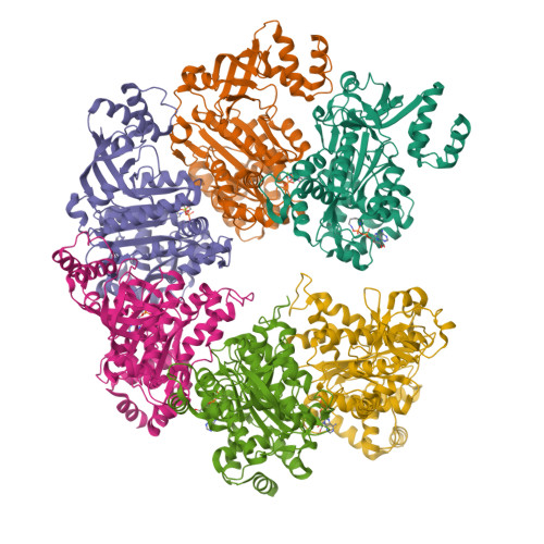
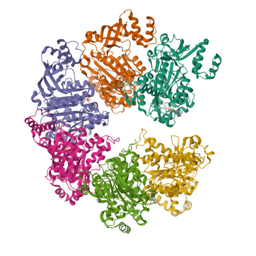
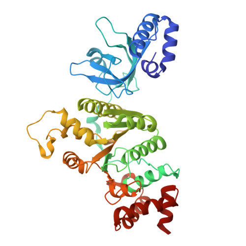
8Q3N

Bacterial transcription termination factor Rho + ADP


Experimental Data Snapshot

  • Method: ELECTRON MICROSCOPY
  • Resolution: 2.63 Å
  • Aggregation State: PARTICLE 
  • Reconstruction Method: SINGLE PARTICLE 

wwPDB Validation   3D Report Full Report


This is version 1.1 of the entry. See complete history


Literature

Transcription termination factor rho polymerizes under stress.

Wang, B.Said, N.Hilal, T.Finazzo, M.Wahl, M.C.Artsimovitch, I.

(2023) bioRxiv 

  • DOI: https://doi.org/10.1101/2023.08.18.553922
  • Primary Citation of Related Structures:  
    8Q3N, 8Q3O, 8Q3P, 8Q3Q

  • PubMed Abstract: 

    Bacterial RNA helicase ρ is a genome sentinel that terminates synthesis of damaged and junk RNAs that are not translated by the ribosome. Co-transcriptional RNA surveillance by ρ is essential for quality control of the transcriptome during optimal growth. However, it is unclear how bacteria protect their RNAs from overzealous ρ during dormancy or stress, conditions common in natural habitats. Here we used cryogenic electron microscopy, biochemical, and genetic approaches to show that residue substitutions, ADP, or ppGpp promote hyper-oligomerization of Escherichia coli ρ. Our results demonstrate that nucleotides bound at subunit interfaces control ρ switching from active hexamers to inactive higher-order oligomers and extended filaments. Polymers formed upon exposure to antibiotics or ppGpp disassemble when stress is relieved, thereby directly linking termination activity to cellular physiology. Inactivation of ρ through hyper-oligomerization is a regulatory strategy shared by RNA polymerases, ribosomes, and metabolic enzymes across all life.


  • Organizational Affiliation

    Department of Microbiology and Center for RNA Biology, The Ohio State University, Columbus, OH, USA.


Macromolecules
Find similar proteins by:  (by identity cutoff)  |  3D Structure
Entity ID: 1
MoleculeChains Sequence LengthOrganismDetailsImage
Transcription termination factor Rho
A, B, C, D, E
A, B, C, D, E, F
419Escherichia coliMutation(s): 0 
Gene Names: rhoAC789_1c41660ACN002_3874EL75_4398EL79_4648EL80_4555
EC: 3.6.4
UniProt
Find proteins for P0AG30 (Escherichia coli (strain K12))
Explore P0AG30 
Go to UniProtKB:  P0AG30
Entity Groups  
Sequence Clusters30% Identity50% Identity70% Identity90% Identity95% Identity100% Identity
UniProt GroupP0AG30
Sequence Annotations
Expand
  • Reference Sequence
Small Molecules
Ligands 2 Unique
IDChains Name / Formula / InChI Key2D Diagram3D Interactions
ADP (Subject of Investigation/LOI)
Query on ADP

Download Ideal Coordinates CCD File 
G [auth A]
H [auth B]
J [auth C]
L [auth D]
N [auth E]
G [auth A],
H [auth B],
J [auth C],
L [auth D],
N [auth E],
P [auth F]
ADENOSINE-5'-DIPHOSPHATE
C10 H15 N5 O10 P2
XTWYTFMLZFPYCI-KQYNXXCUSA-N
MG
Query on MG

Download Ideal Coordinates CCD File 
I [auth B],
K [auth C],
M [auth D],
O [auth E]
MAGNESIUM ION
Mg
JLVVSXFLKOJNIY-UHFFFAOYSA-N
Experimental Data & Validation

Experimental Data

  • Method: ELECTRON MICROSCOPY
  • Resolution: 2.63 Å
  • Aggregation State: PARTICLE 
  • Reconstruction Method: SINGLE PARTICLE 
EM Software:
TaskSoftware PackageVersion
RECONSTRUCTIONcryoSPARCv3.3.2

Structure Validation

View Full Validation Report


Entry History & Funding Information

Deposition Data


Funding OrganizationLocationGrant Number
German Research Foundation (DFG)Germany433623608

Revision History  (Full details and data files)

  • Version 1.0: 2023-09-27
    Type: Initial release
  • Version 1.1: 2023-10-04
    Changes: Advisory, Derived calculations