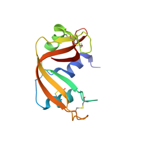
Experimental phasing opportunities for macromolecular crystallography at very long wavelengths.
El Omari, K., Duman, R., Mykhaylyk, V., Orr, C.M., Latimer-Smith, M., Winter, G., Grama, V., Qu, F., Bountra, K., Kwong, H.S., Romano, M., Reis, R.I., Vogeley, L., Vecchia, L., Owen, C.D., Wittmann, S., Renner, M., Senda, M., Matsugaki, N., Kawano, Y., Bowden, T.A., Moraes, I., Grimes, J.M., Mancini, E.J., Walsh, M.A., Guzzo, C.R., Owens, R.J., Jones, E.Y., Brown, D.G., Stuart, D.I., Beis, K., Wagner, A.(2023) Commun Chem 6: 219-219
- PubMed: 37828292
- DOI: https://doi.org/10.1038/s42004-023-01014-0
- Primary Citation of Related Structures:
8PWN, 8PX0, 8PX1, 8PX4, 8PX5, 8PX7, 8PX9, 8PXC, 8PXG, 8PXH, 8PXJ, 8PXK, 8PXL, 8PYV, 8PYZ, 8PZ4, 8PZ5 - PubMed Abstract:
Despite recent advances in cryo-electron microscopy and artificial intelligence-based model predictions, a significant fraction of structure determinations by macromolecular crystallography still requires experimental phasing, usually by means of single-wavelength anomalous diffraction (SAD) techniques. Most synchrotron beamlines provide highly brilliant beams of X-rays of between 0.7 and 2 Å wavelength. Use of longer wavelengths to access the absorption edges of biologically important lighter atoms such as calcium, potassium, chlorine, sulfur and phosphorus for native-SAD phasing is attractive but technically highly challenging. The long-wavelength beamline I23 at Diamond Light Source overcomes these limitations and extends the accessible wavelength range to λ = 5.9 Å. Here we report 22 macromolecular structures solved in this extended wavelength range, using anomalous scattering from a range of elements which demonstrate the routine feasibility of lighter atom phasing. We suggest that, in light of its advantages, long-wavelength crystallography is a compelling option for experimental phasing.
Organizational Affiliation:
Diamond Light Source, Harwell Science and Innovation Campus, -, OX110DE, UK.


