8PVB | pdb_00008pvb

Structure of GABAAR determined by cryoEM at 100 keV


Experimental Data Snapshot

  • Method: ELECTRON MICROSCOPY
  • Resolution: 3.60 Å
  • Aggregation State: PARTICLE 
  • Reconstruction Method: SINGLE PARTICLE 

Starting Model: experimental
View more details

wwPDB Validation 3D Report Full Report

Validation slider image for 8PVB

This is version 1.3 of the entry. See complete history

Literature

Structure determination by cryoEM at 100 keV.

McMullan, G.Naydenova, K.Mihaylov, D.Yamashita, K.Peet, M.J.Wilson, H.Dickerson, J.L.Chen, S.Cannone, G.Lee, Y.Hutchings, K.A.Gittins, O.Sobhy, M.A.Wells, T.El-Gomati, M.M.Dalby, J.Meffert, M.Schulze-Briese, C.Henderson, R.Russo, C.J.

(2023) Proc Natl Acad Sci U S A 120: e2312905120-e2312905120

  • DOI: https://doi.org/10.1073/pnas.2312905120
  • Primary Citation Related Structures: 
    8PV9, 8PVA, 8PVB, 8PVC, 8PVD, 8PVE, 8PVF, 8PVG, 8PVH, 8PVI, 8PVJ

  • PubMed Abstract: 

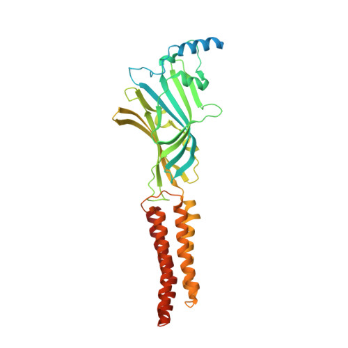
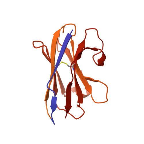
    Electron cryomicroscopy can, in principle, determine the structures of most biological molecules but is currently limited by access, specimen preparation difficulties, and cost. We describe a purpose-built instrument operating at 100 keV-including advances in electron optics, detection, and processing-that makes structure determination fast and simple at a fraction of current costs. The instrument attains its theoretical performance limits, allowing atomic resolution imaging of gold test specimens and biological molecular structure determination in hours. We demonstrate its capabilities by determining the structures of eleven different specimens, ranging in size from 140 kDa to 2 MDa, using a fraction of the data normally required. CryoEM with a microscope designed specifically for high-efficiency, on-the-spot imaging of biological molecules will expand structural biology to a wide range of previously intractable problems.


  • Organizational Affiliation
    • Medical Research Council (MRC) Laboratory of Molecular Biology, Cambridge CB2 0QH, United Kingdom.

Macromolecule Content 

  • Total Structure Weight: 104.6 kDa 
  • Atom Count: 3,845 
  • Modeled Residue Count: 455 
  • Deposited Residue Count: 918 
  • Unique protein chains: 2

Macromolecules

Find similar proteins by:|  3D Structure
Entity ID: 1
MoleculeChains  Sequence LengthOrganismDetailsImage
Gamma-aminobutyric acid receptor subunit beta-3396Homo sapiensMutation(s): 0 
Gene Names: GABRB3
Membrane Entity: Yes 
UniProt & NIH Common Fund Data Resources
Find proteins for P28472 (Homo sapiens)
Explore P28472 
Go to UniProtKB:  P28472
PHAROS:  P28472
GTEx:  ENSG00000166206 
Entity Groups
Sequence Clusters30% Identity50% Identity70% Identity90% Identity95% Identity100% Identity
UniProt GroupP28472
Glycosylation
Glycosylation Sites: 2Go to GlyGen: P28472-1
Sequence Annotations
Expand
Reference Sequence
Find similar proteins by:|  3D Structure
Entity ID: 2
MoleculeChains  Sequence LengthOrganismDetailsImage
Megabody Mb25B [auth O]522Lama glamaMutation(s): 0 

Oligosaccharides

Help  
Entity ID: 3
MoleculeChains Length2D Diagram GlycosylationD Interactions
2-acetamido-2-deoxy-beta-D-glucopyranose-(1-4)-2-acetamido-2-deoxy-beta-D-glucopyranoseC [auth B]2N-Glycosylation
Glycosylation Resources
GlyTouCan: G42666HT
GlyCosmos: G42666HT
GlyGen: G42666HT
Entity ID: 4
MoleculeChains Length2D Diagram GlycosylationD Interactions
alpha-D-mannopyranose-(1-3)-[alpha-D-mannopyranose-(1-6)]beta-D-mannopyranose-(1-4)-2-acetamido-2-deoxy-beta-D-glucopyranose-(1-4)-2-acetamido-2-deoxy-beta-D-glucopyranoseD [auth C]5N-Glycosylation
Glycosylation Resources
GlyTouCan: G22768VO
GlyCosmos: G22768VO
GlyGen: G22768VO

Small Molecules

Ligands 5 Unique
IDChains Name / Formula / InChI Key2D Diagram3D Interactions
V8D

Query on V8D


Download:Ideal Coordinates CCD File
M [auth A]Etomidate
C14 H16 N2 O2
NPUKDXXFDDZOKR-LLVKDONJSA-N
R16

Query on R16


Download:Ideal Coordinates CCD File
E [auth A]HEXADECANE
C16 H34
DCAYPVUWAIABOU-UHFFFAOYSA-N
D10

Query on D10


Download:Ideal Coordinates CCD File
F [auth A],
G [auth A],
H [auth A],
I [auth A]
DECANE
C10 H22
DIOQZVSQGTUSAI-UHFFFAOYSA-N
HSM

Query on HSM


Download:Ideal Coordinates CCD File
J [auth A]HISTAMINE
C5 H9 N3
NTYJJOPFIAHURM-UHFFFAOYSA-N
CL

Query on CL


Download:Ideal Coordinates CCD File
K [auth A],
L [auth A]
CHLORIDE ION
Cl
VEXZGXHMUGYJMC-UHFFFAOYSA-M
Binding Affinity Annotations 
IDSourceBinding Affinity
V8D BindingDB:  8PVB IC50: 5.77e+5 (nM) from 1 assay(s)
EC50: min: 1200, max: 6.40e+4 (nM) from 4 assay(s)

Experimental Data & Validation

Experimental Data

  • Method: ELECTRON MICROSCOPY
  • Resolution: 3.60 Å
  • Aggregation State: PARTICLE 
  • Reconstruction Method: SINGLE PARTICLE 
EM Software:
TaskSoftware PackageVersion
MODEL REFINEMENTServalcat0.4.27

Structure Validation

View Full Validation Report


Entry History 

& Funding Information

Deposition Data


Funding OrganizationLocationGrant Number
Medical Research Council (MRC, United Kingdom)United KingdomMC UP 120117
Medical Research Council (MRC, United Kingdom)United KingdomMC U105184322
Wellcome TrustUnited Kingdom220526/B/20/Z
Engineering and Physical Sciences Research CouncilUnited KingdomR122522
Innovate UKUnited Kingdom103806
Biotechnology and Biological Sciences Research Council (BBSRC)United KingdomBB/T003677/1

Revision History  (Full details and data files)

  • Version 1.0: 2023-11-29
    Type: Initial release
  • Version 1.1: 2023-12-06
    Changes: Database references
  • Version 1.2: 2024-01-31
    Changes: Derived calculations, Structure summary
  • Version 1.3: 2024-10-09
    Changes: Data collection, Structure summary