8PUO | pdb_00008puo

Structural determinants of cold-activity and glucose tolerance of a family 1 glycoside hydrolase (GH1) from Antarctic Marinomonas Ef 1


Experimental Data Snapshot

  • Method: X-RAY DIFFRACTION
  • Resolution: 1.80 Å
  • R-Value Free: 
    0.189 (Depositor), 0.193 (DCC) 
  • R-Value Work: 
    0.165 (Depositor), 0.170 (DCC) 
  • R-Value Observed: 
    0.165 (Depositor) 

Starting Model: experimental
View more details

wwPDB Validation 3D Report Full Report

Validation slider image for 8PUO

This is version 1.1 of the entry. See complete history

Literature

Structural determinants of cold activity and glucose tolerance of a family 1 glycoside hydrolase (GH1) from Antarctic Marinomonas sp. ef1.

Gourlay, L.J.Mangiagalli, M.Moroni, E.Lotti, M.Nardini, M.

(2024) FEBS J 291: 2897-2917

  • DOI: https://doi.org/10.1111/febs.17096
  • Primary Citation Related Structures: 
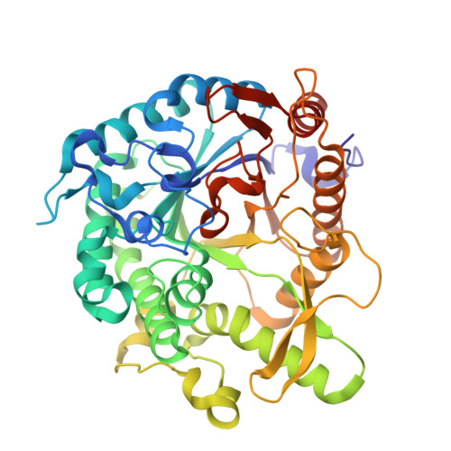
    8PUO

  • PubMed Abstract: 

    Cold-active enzymes support life at low temperatures due to their ability to maintain high activity in the cold and can be useful in several biotechnological applications. Although information on the mechanisms of enzyme cold adaptation is still too limited to devise general rules, it appears that very diverse structural and functional changes are exploited in different protein families and within the same family. In this context, we studied the cold adaptation mechanism and the functional properties of a member of the glycoside hydrolase family 1 (GH1) from the Antarctic bacterium Marinomonas sp. ef1. This enzyme exhibits all typical functional hallmarks of cold adaptation, including high catalytic activity at 5 °C, broad substrate specificity, low thermal stability, and higher lability of the active site compared to the overall structure. Analysis of the here-reported crystal structure (1.8 Å resolution) and molecular dynamics simulations suggest that cold activity and thermolability may be due to a flexible region around the active site (residues 298-331), whereas the dynamic behavior of loops flanking the active site (residues 47-61 and 407-413) may favor enzyme-substrate interactions at the optimal temperature of catalysis (T opt ) by tethering together protein regions lining the active site. Stapling of the N-terminus onto the surface of the β-barrel is suggested to partly counterbalance protein flexibility, thus providing a stabilizing effect. The tolerance of the enzyme to glucose and galactose is accounted for by the presence of a "gatekeeping" hydrophobic residue (Leu178), located at the entrance of the active site.


  • Organizational Affiliation
    • Department of Biosciences, University of Milano, Italy.

Macromolecule Content 

  • Total Structure Weight: 105.36 kDa 
  • Atom Count: 7,862 
  • Modeled Residue Count: 889 
  • Deposited Residue Count: 910 
  • Unique protein chains: 1

Macromolecules

Find similar proteins by:|  3D Structure
Entity ID: 1
MoleculeChains  Sequence LengthOrganismDetailsImage
Beta-glucosidase
A, B
455Marinomonas sp. ef1Mutation(s): 0 
Gene Names: TY87_18135
EC: 3.2.1.21
UniProt
Find proteins for A0ACD6BA54 (Marinomonas sp. BSi20584)
Explore A0ACD6BA54 
Go to UniProtKB:  A0ACD6BA54
Entity Groups
Sequence Clusters30% Identity50% Identity70% Identity90% Identity95% Identity100% Identity
UniProt GroupA0ACD6BA54
Sequence Annotations
Expand
Reference Sequence

Small Molecules

Ligands 2 Unique
IDChains Name / Formula / InChI Key2D Diagram3D Interactions
TRS

Query on TRS


Download:Ideal Coordinates CCD File
BA [auth B],
O [auth A]
2-AMINO-2-HYDROXYMETHYL-PROPANE-1,3-DIOL
C4 H12 N O3
LENZDBCJOHFCAS-UHFFFAOYSA-O
EDO

Query on EDO


Download:Ideal Coordinates CCD File
AA [auth B]
C [auth A]
D [auth A]
E [auth A]
F [auth A]
AA [auth B],
C [auth A],
D [auth A],
E [auth A],
F [auth A],
G [auth A],
H [auth A],
I [auth A],
J [auth A],
K [auth A],
L [auth A],
M [auth A],
N [auth A],
P [auth B],
Q [auth B],
R [auth B],
S [auth B],
T [auth B],
U [auth B],
V [auth B],
W [auth B],
X [auth B],
Y [auth B],
Z [auth B]
1,2-ETHANEDIOL
C2 H6 O2
LYCAIKOWRPUZTN-UHFFFAOYSA-N

Experimental Data & Validation

Experimental Data

  • Method: X-RAY DIFFRACTION
  • Resolution: 1.80 Å
  • R-Value Free:  0.189 (Depositor), 0.193 (DCC) 
  • R-Value Work:  0.165 (Depositor), 0.170 (DCC) 
  • R-Value Observed: 0.165 (Depositor) 
Space Group: P 21 21 2
Unit Cell:
Length ( Å )Angle ( ˚ )
a = 103.116α = 90
b = 201.12β = 90
c = 47.825γ = 90
Software Package:
Software NamePurpose
PHENIXrefinement
XDSdata reduction
Aimlessdata scaling
MOLREPphasing

Structure Validation

View Full Validation Report


Entry History 

& Funding Information

Deposition Data


Funding OrganizationLocationGrant Number
Not funded--

Revision History  (Full details and data files)

  • Version 1.0: 2024-03-06
    Type: Initial release
  • Version 1.1: 2024-07-17
    Changes: Database references