8PNV | pdb_00008pnv

Cryo-EM structure of styrene oxide isomerase


Experimental Data Snapshot

  • Method: ELECTRON MICROSCOPY
  • Resolution: 2.05 Å
  • Aggregation State: PARTICLE 
  • Reconstruction Method: SINGLE PARTICLE 

wwPDB Validation 3D Report Full Report

Validation slider image for 8PNV

This is version 1.4 of the entry. See complete history

Literature

Structural basis of the Meinwald rearrangement catalysed by styrene oxide isomerase.

Khanppnavar, B.Choo, J.P.S.Hagedoorn, P.L.Smolentsev, G.Stefanic, S.Kumaran, S.Tischler, D.Winkler, F.K.Korkhov, V.M.Li, Z.Kammerer, R.A.Li, X.

(2024) Nat Chem 16: 1496-1504

  • DOI: https://doi.org/10.1038/s41557-024-01523-y
  • Primary Citation Related Structures: 
    8PNU, 8PNV

  • PubMed Abstract: 

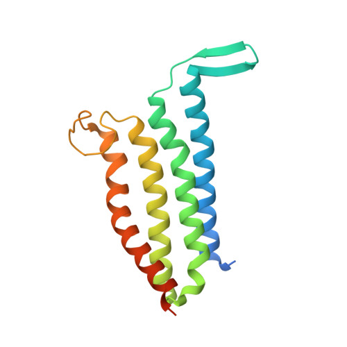
    Membrane-bound styrene oxide isomerase (SOI) catalyses the Meinwald rearrangement-a Lewis-acid-catalysed isomerization of an epoxide to a carbonyl compound-and has been used in single and cascade reactions. However, the structural information that explains its reaction mechanism has remained elusive. Here we determine cryo-electron microscopy (cryo-EM) structures of SOI bound to a single-domain antibody with and without the competitive inhibitor benzylamine, and elucidate the catalytic mechanism using electron paramagnetic resonance spectroscopy, functional assays, biophysical methods and docking experiments. We find ferric haem b bound at the subunit interface of the trimeric enzyme through H58, where Fe(III) acts as the Lewis acid by binding to the epoxide oxygen. Y103 and N64 and a hydrophobic pocket binding the oxygen of the epoxide and the aryl group, respectively, position substrates in a manner that explains the high regio-selectivity and stereo-specificity of SOI. Our findings can support extending the range of epoxide substrates and be used to potentially repurpose SOI for the catalysis of new-to-nature Fe-based chemical reactions.


  • Organizational Affiliation
    • Laboratory of Biomolecular Research, Division of Biology and Chemistry, Paul Scherrer Institute, Villigen, Switzerland.

Macromolecule Content 

  • Total Structure Weight: 206.12 kDa 
  • Atom Count: 13,098 
  • Modeled Residue Count: 1,626 
  • Deposited Residue Count: 1,872 
  • Unique protein chains: 2

Macromolecules

Find similar proteins by:|  3D Structure
Entity ID: 1
MoleculeChains  Sequence LengthOrganismDetailsImage
Styrene oxide isomerase
A, B, C, G, H
A, B, C, G, H, I
183Pseudomonas sp. VLB120Mutation(s): 0 
Gene Names: stdC
Membrane Entity: Yes 
UniProt
Find proteins for O50216 (Pseudomonas sp. VLB120)
Explore O50216 
Go to UniProtKB:  O50216
Entity Groups
Sequence Clusters30% Identity50% Identity70% Identity90% Identity95% Identity100% Identity
UniProt GroupO50216
Sequence Annotations
Expand
Reference Sequence
Find similar proteins by:|  3D Structure
Entity ID: 2
MoleculeChains  Sequence LengthOrganismDetailsImage
Nanobody
D, E, F, J, K
D, E, F, J, K, L
129Vicugna pacosMutation(s): 0 

Experimental Data & Validation

Experimental Data

  • Method: ELECTRON MICROSCOPY
  • Resolution: 2.05 Å
  • Aggregation State: PARTICLE 
  • Reconstruction Method: SINGLE PARTICLE 
EM Software:
TaskSoftware PackageVersion
MODEL REFINEMENTPHENIX
RECONSTRUCTIONRELION3.1.3

Structure Validation

View Full Validation Report


Entry History 

& Funding Information

Deposition Data


Funding OrganizationLocationGrant Number
Not funded--

Revision History  (Full details and data files)

  • Version 1.0: 2024-04-03
    Type: Initial release
  • Version 1.1: 2024-06-12
    Changes: Database references
  • Version 1.2: 2024-09-18
    Changes: Data collection, Database references
  • Version 1.3: 2024-10-16
    Changes: Data collection, Structure summary
  • Version 1.4: 2025-07-02
    Changes: Data collection