8PKE | pdb_00008pke

ATTRV20I amyloid fibril from hereditary ATTR amloidosis


Experimental Data Snapshot

  • Method: ELECTRON MICROSCOPY
  • Resolution: 3.39 Å
  • Aggregation State: HELICAL ARRAY 
  • Reconstruction Method: HELICAL 

wwPDB Validation 3D Report Full Report

Validation slider image for 8PKE

This is version 1.0 of the entry. See complete history

Literature

Common transthyretin-derived amyloid fibril structures in patients with hereditary ATTR amyloidosis.

Steinebrei, M.Baur, J.Pradhan, A.Kupfer, N.Wiese, S.Hegenbart, U.Schonland, S.O.Schmidt, M.Fandrich, M.

(2023) Nat Commun 14: 7623-7623

  • DOI: https://doi.org/10.1038/s41467-023-43301-3
  • Primary Citation Related Structures: 
    8PKE, 8PKF, 8PKG

  • PubMed Abstract: 

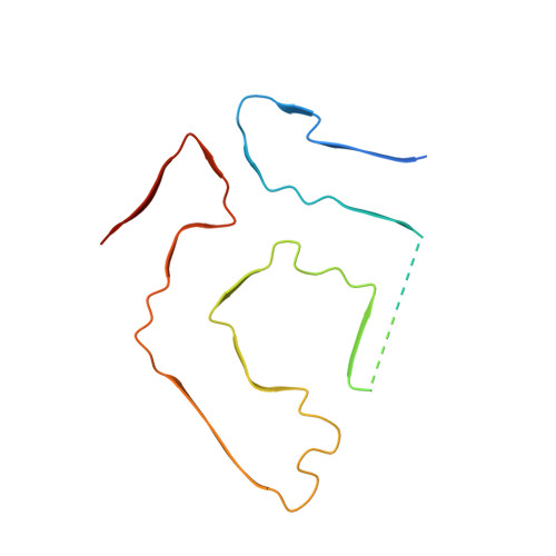
    Systemic ATTR amyloidosis is an increasingly important protein misfolding disease that is provoked by the formation of amyloid fibrils from transthyretin protein. The pathological and clinical disease manifestations and the number of pathogenic mutational changes in transthyretin are highly diverse, raising the question whether the different mutations may lead to different fibril morphologies. Using cryo-electron microscopy, however, we show here that the fibril structure is remarkably similar in patients that are affected by different mutations. Our data suggest that the circumstances under which these fibrils are formed and deposited inside the body - and not only the fibril morphology - are crucial for defining the phenotypic variability in many patients.


  • Organizational Affiliation
    • Institute of Protein Biochemistry, Ulm University, Helmholtzstrasse 8/1, Ulm, D-89081, Germany. maximilian.steinebrei@uni-ulm.de.

Macromolecule Content 

  • Total Structure Weight: 82.75 kDa 
  • Atom Count: 4,314 
  • Modeled Residue Count: 552 
  • Deposited Residue Count: 762 
  • Unique protein chains: 1

Macromolecules

Find similar proteins by:|  3D Structure
Entity ID: 1
MoleculeChains  Sequence LengthOrganismDetailsImage
Transthyretin
A, B, C, D, E
A, B, C, D, E, F
127Homo sapiensMutation(s): 1 
UniProt & NIH Common Fund Data Resources
Find proteins for P02766 (Homo sapiens)
Explore P02766 
Go to UniProtKB:  P02766
PHAROS:  P02766
GTEx:  ENSG00000118271 
Entity Groups
Sequence Clusters30% Identity50% Identity70% Identity90% Identity95% Identity100% Identity
UniProt GroupP02766
Sequence Annotations
Expand
Reference Sequence

Experimental Data & Validation

Experimental Data

  • Method: ELECTRON MICROSCOPY
  • Resolution: 3.39 Å
  • Aggregation State: HELICAL ARRAY 
  • Reconstruction Method: HELICAL 
EM Software:
TaskSoftware PackageVersion
RECONSTRUCTIONPHENIX

Structure Validation

View Full Validation Report


Entry History 

& Funding Information

Deposition Data


Funding OrganizationLocationGrant Number
German Research Foundation (DFG)GermanyFA 456/28

Revision History  (Full details and data files)

  • Version 1.0: 2023-12-06
    Type: Initial release