8PJB | pdb_00008pjb

Cryo-EM structure of MLE in complex with UUC RNA and ADP


Experimental Data Snapshot

  • Method: ELECTRON MICROSCOPY
  • Resolution: 3.62 Å
  • Aggregation State: PARTICLE 
  • Reconstruction Method: SINGLE PARTICLE 

wwPDB Validation 3D Report Full Report

Validation slider image for 8PJB

This is version 1.2 of the entry. See complete history

Literature

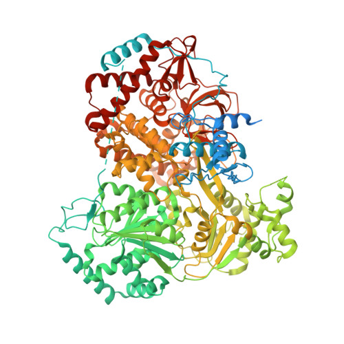
Structural basis of RNA-induced autoregulation of the DExH-type RNA helicase maleless.

Jagtap, P.K.A.Muller, M.Kiss, A.E.Thomae, A.W.Lapouge, K.Beck, M.Becker, P.B.Hennig, J.

(2023) Mol Cell 83: 4318-4333.e10

  • DOI: https://doi.org/10.1016/j.molcel.2023.10.026
  • Primary Citation Related Structures: 
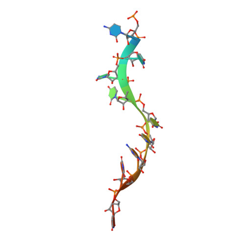
    8B9G, 8B9I, 8B9J, 8B9K, 8B9L, 8PJB, 8PJJ

  • PubMed Abstract: 

    RNA unwinding by DExH-type helicases underlies most RNA metabolism and function. It remains unresolved if and how the basic unwinding reaction of helicases is regulated by auxiliary domains. We explored the interplay between the RecA and auxiliary domains of the RNA helicase maleless (MLE) from Drosophila using structural and functional studies. We discovered that MLE exists in a dsRNA-bound open conformation and that the auxiliary dsRBD2 domain aligns the substrate RNA with the accessible helicase tunnel. In an ATP-dependent manner, dsRBD2 associates with the helicase module, leading to tunnel closure around ssRNA. Furthermore, our structures provide a rationale for blunt-ended dsRNA unwinding and 3'-5' translocation by MLE. Structure-based MLE mutations confirm the functional relevance of our model for RNA unwinding. Our findings contribute to our understanding of the fundamental mechanics of auxiliary domains in DExH helicase MLE, which serves as a model for its human ortholog and potential therapeutic target, DHX9/RHA.


  • Organizational Affiliation
    • Structural and Computational Biology Unit, EMBL Heidelberg, Meyerhofstraße 1, 69117 Heidelberg, Germany; Chair of Biochemistry IV, Biophysical Chemistry, University of Bayreuth, Bayreuth, Germany. Electronic address: pravin.jagtap@embl.de.

Macromolecule Content 

  • Total Structure Weight: 134.41 kDa 
  • Atom Count: 7,840 
  • Modeled Residue Count: 971 
  • Deposited Residue Count: 1,170 
  • Unique protein chains: 1
  • Unique nucleic acid chains: 1

Macromolecules


Find similar proteins by:|  3D Structure
Entity ID: 1
MoleculeChains  Sequence LengthOrganismDetailsImage
Dosage compensation regulator1,158Drosophila melanogasterMutation(s): 0 
Gene Names: mlenapCG11680
EC: 3.6.4.13
UniProt
Find proteins for P24785 (Drosophila melanogaster)
Explore P24785 
Go to UniProtKB:  P24785
Entity Groups
Sequence Clusters30% Identity50% Identity70% Identity90% Identity95% Identity100% Identity
UniProt GroupP24785
Sequence Annotations
Expand
Reference Sequence
Find similar nucleic acids by:  Sequence
Entity ID: 2
MoleculeChains LengthOrganismImage
RNA (5'-R(P*CP*UP*CP*UP*UP*UP*CP*UP*U)-3')B [auth C]12Drosophila melanogaster
Sequence Annotations
Expand
Reference Sequence

Experimental Data & Validation

Experimental Data

  • Method: ELECTRON MICROSCOPY
  • Resolution: 3.62 Å
  • Aggregation State: PARTICLE 
  • Reconstruction Method: SINGLE PARTICLE 
EM Software:
TaskSoftware PackageVersion
MODEL REFINEMENTPHENIX

Structure Validation

View Full Validation Report


Entry History 

& Funding Information

Deposition Data


Funding OrganizationLocationGrant Number
EIPOD fellowship under Marie Sklodowska-Curie Actions COFUNDGermany--

Revision History  (Full details and data files)

  • Version 1.0: 2023-11-01
    Type: Initial release
  • Version 1.1: 2024-06-05
    Changes: Database references
  • Version 1.2: 2025-07-02
    Changes: Data collection, Structure summary