8PDS

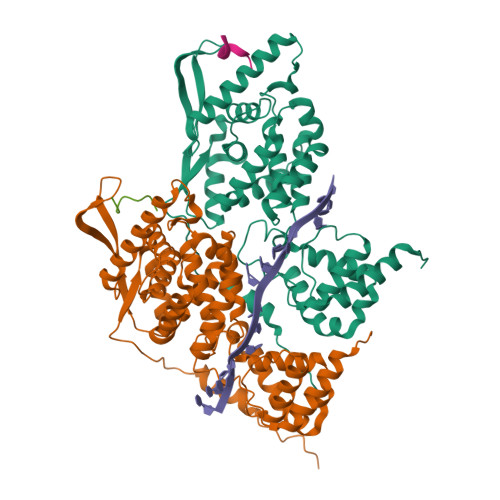
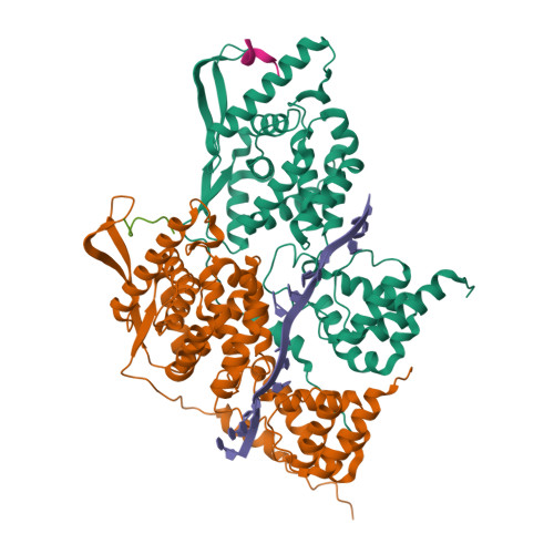
Local refinement of dimeric HMPV N-RNA bound to the C-terminal region of P


Experimental Data Snapshot

  • Method: ELECTRON MICROSCOPY
  • Resolution: 2.90 Å
  • Aggregation State: PARTICLE 
  • Reconstruction Method: SINGLE PARTICLE 

wwPDB Validation   3D Report Full Report


This is version 1.1 of the entry. See complete history


Literature

Structure of the N-RNA/P interface indicates mode of L/P recruitment to the nucleocapsid of human metapneumovirus.

Whitehead, J.D.Decool, H.Leyrat, C.Carrique, L.Fix, J.Eleouet, J.F.Galloux, M.Renner, M.

(2023) Nat Commun 14: 7627-7627

  • DOI: https://doi.org/10.1038/s41467-023-43434-5
  • Primary Citation of Related Structures:  
    8PDL, 8PDM, 8PDN, 8PDO, 8PDP, 8PDQ, 8PDR, 8PDS

  • PubMed Abstract: 

    Human metapneumovirus (HMPV) is a major cause of respiratory illness in young children. The HMPV polymerase (L) binds an obligate cofactor, the phosphoprotein (P). During replication and transcription, the L/P complex traverses the viral RNA genome, which is encapsidated within nucleoproteins (N). An essential interaction between N and a C-terminal region of P tethers the L/P polymerase to the template. This N-P interaction is also involved in the formation of cytoplasmic viral factories in infected cells, called inclusion bodies. To define how the polymerase component P recognizes N-encapsidated RNA (N-RNA) we employed cryogenic electron microscopy (cryo-EM) and molecular dynamics simulations, coupled to activity assays and imaging of inclusion bodies in cells. We report a 2.9 Å resolution structure of a triple-complex between multimeric N, bound to both RNA and the C-terminal region of P. Furthermore, we also present cryo-EM structures of assembled N in different oligomeric states, highlighting the plasticity of N. Combined with our functional assays, these structural data delineate in molecular detail how P attaches to N-RNA whilst retaining substantial conformational dynamics. Moreover, the N-RNA-P triple complex structure provides a molecular blueprint for the design of therapeutics to potentially disrupt the attachment of L/P to its template.


  • Organizational Affiliation

    Division of Structural Biology, The Wellcome Centre for Human Genetics, University of Oxford, Oxford, UK.


Macromolecules

Find similar proteins by:  (by identity cutoff)  |  3D Structure
Entity ID: 1
MoleculeChains Sequence LengthOrganismDetailsImage
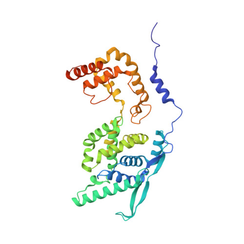
NucleoproteinA,
D [auth C]
394Human metapneumovirus CAN97-83Mutation(s): 2 
Gene Names: N
UniProt
Find proteins for Q6WBA1 (Human metapneumovirus (strain CAN97-83))
Explore Q6WBA1 
Go to UniProtKB:  Q6WBA1
Entity Groups  
Sequence Clusters30% Identity50% Identity70% Identity90% Identity95% Identity100% Identity
UniProt GroupQ6WBA1
Sequence Annotations
Expand
  • Reference Sequence

Find similar proteins by:  Sequence   |   3D Structure  

Entity ID: 3
MoleculeChains Sequence LengthOrganismDetailsImage
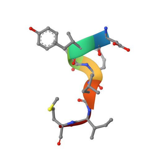
PhosphoproteinC [auth B],
E [auth D]
7Human metapneumovirus CAN97-83Mutation(s): 0 
UniProt
Find proteins for Q8B9Q8 (Human metapneumovirus (strain CAN97-83))
Explore Q8B9Q8 
Go to UniProtKB:  Q8B9Q8
Entity Groups  
UniProt GroupQ8B9Q8
Sequence Annotations
Expand
  • Reference Sequence

Find similar nucleic acids by:  Sequence   |   3D Structure  

Entity ID: 2
MoleculeChains LengthOrganismImage
RNAB [auth E]14Escherichia coli
Sequence Annotations
Expand
  • Reference Sequence
Experimental Data & Validation

Experimental Data

  • Method: ELECTRON MICROSCOPY
  • Resolution: 2.90 Å
  • Aggregation State: PARTICLE 
  • Reconstruction Method: SINGLE PARTICLE 
EM Software:
TaskSoftware PackageVersion
MODEL REFINEMENTPHENIX
RECONSTRUCTIONcryoSPARC

Structure Validation

View Full Validation Report


Entry History & Funding Information

Deposition Data


Funding OrganizationLocationGrant Number
Wellcome TrustUnited Kingdom--

Revision History  (Full details and data files)

  • Version 1.0: 2023-12-06
    Type: Initial release
  • Version 1.1: 2024-03-20
    Changes: Database references, Source and taxonomy, Structure summary