Discovery of BAY-405: An Azaindole-Based MAP4K1 Inhibitor for the Enhancement of T-Cell Immunity against Cancer.
Mowat, J., Carretero, R., Leder, G., Aiguabella Font, N., Neuhaus, R., Berndt, S., Gunther, J., Friberg, A., Schafer, M., Briem, H., Raschke, M., Miyatake Ondozabal, H., Buchmann, B., Boemer, U., Kreft, B., Hartung, I.V., Offringa, R.(2024) J Med Chem 67: 17429-17453
- PubMed: 39331123 Search on PubMedSearch on PubMed Central
- DOI: https://doi.org/10.1021/acs.jmedchem.4c01325
- Primary Citation Related Structures:
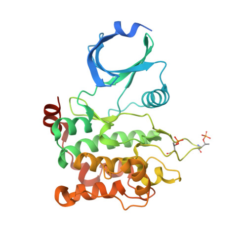
8PAR, 8PAS, 8PAU, 8PAV, 8PAW - PubMed Abstract:
Mitogen-activated protein kinase kinase kinase kinase 1 (MAP4K1) is a serine/threonine kinase that acts as an immune checkpoint downstream of T-cell receptor stimulation. MAP4K1 activity is enhanced by prostaglandin E2 (PGE2) and transforming growth factor beta (TGFβ), immune modulators commonly present in the tumor microenvironment. Therefore, its pharmacological inhibition is an attractive immuno-oncology concept for inducing therapeutic T-cell responses in cancer patients. Here, we describe the systematic optimization of azaindole-based lead compound 1 , resulting in the discovery of potent and selective MAP4K1 inhibitor 38 (BAY-405) that displays nanomolar potency in biochemical and cellular assays as well as in vivo exposure after oral dosing. BAY-405 enhances T-cell immunity and overcomes the suppressive effect of PGE2 and TGFβ. Treatment of tumor-bearing mice shows T-cell-dependent antitumor efficacy. MAP4K1 inhibition in conjunction with PD-L1 blockade results in a superior antitumor impact, illustrating the complementarity of the single agent treatments.
- Bayer AG, Pharmaceutical R&D, 13342 Berlin, Germany.
Organizational Affiliation: