8P8E | pdb_00008p8e

Crystal structure of endolysin gp46 from Pseudomonas aeruginosa bacteriophage vB_PaeM_KTN6


Experimental Data Snapshot

  • Method: X-RAY DIFFRACTION
  • Resolution: 1.39 Å
  • R-Value Free: 
    0.164 (Depositor), 0.164 (DCC) 
  • R-Value Work: 
    0.126 (Depositor), 0.126 (DCC) 

Starting Model: experimental
View more details

wwPDB Validation 3D Report Full Report

Validation slider image for 8P8E

This is version 1.1 of the entry. See complete history

Literature

Structural and Biochemical Characterization of a New Phage-Encoded Muramidase, KTN6 Gp46.

Sanz-Gaitero, M.De Maesschalck, V.Patel, A.Longin, H.Van Noort, V.Rodriguez-Rubio, L.van Ryne, M.Danis-Wlodarczyk, K.Drulis-Kawa, Z.Mesnage, S.van Raaij, M.Lavigne, R.

(2024) Phage (New Rochelle) 5: 53-62

  • DOI: https://doi.org/10.1089/phage.2023.0040
  • Primary Citation Related Structures: 
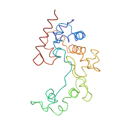
    8P8E

  • PubMed Abstract: 

    Endolysins are phage-encoded lytic enzymes that degrade bacterial peptidoglycan at the end of phage lytic cycles to release new phage particles. These enzymes are being explored as an alternative to small-molecule antibiotics. The crystal structure of KTN6 Gp46 was determined and compared with a ColabFold model. Cleavage specificity was examined using a peptidoglycan digest and reversed-phase high-performance liquid chromatography coupled to mass spectrometry (HPLC/MS). The structure of KTN6 Gp46 could be determined at 1.4 Å resolution, and key differences in loops of the putative peptidoglycan binding domain were identified in comparison with its closest known homologue, the endolysin of phage SPN1S. Reversed-phase HPLC/MS analysis of the reaction products following peptidoglycan digestion confirmed the muramidase activity of Gp46, consistent with structural predictions. These insights into the structure and function of endolysins further expand the toolbox for endolysin engineering and explore their potential in enzyme-based antibacterial design strategies.


  • Organizational Affiliation
    • Centro Nacional de Biotecnologia, Consejo Superior de Investigaciones Cientificas, Madrid, Spain.

Macromolecule Content 

  • Total Structure Weight: 24.78 kDa 
  • Atom Count: 1,959 
  • Modeled Residue Count: 218 
  • Deposited Residue Count: 220 
  • Unique protein chains: 1

Macromolecules

Find similar proteins by:|  3D Structure
Entity ID: 1
MoleculeChains  Sequence LengthOrganismDetailsImage
Glycoside hydrolase family proteinA [auth AAA]220Bacteriophage sp.Mutation(s): 0 
Gene Names: IDBGMNHM_00003
UniProt
Find proteins for A0ACD6BAN9 (Pseudomonas phage Ka2)
Explore A0ACD6BAN9 
Go to UniProtKB:  A0ACD6BAN9
Entity Groups
Sequence Clusters30% Identity50% Identity70% Identity90% Identity95% Identity100% Identity
UniProt GroupA0ACD6BAN9
Sequence Annotations
Expand
Reference Sequence

Experimental Data & Validation

Experimental Data

  • Method: X-RAY DIFFRACTION
  • Resolution: 1.39 Å
  • R-Value Free:  0.164 (Depositor), 0.164 (DCC) 
  • R-Value Work:  0.126 (Depositor), 0.126 (DCC) 
Space Group: P 21 21 21
Unit Cell:
Length ( Å )Angle ( ˚ )
a = 41.796α = 90
b = 43.201β = 90
c = 126.181γ = 90
Software Package:
Software NamePurpose
REFMACrefinement
XDSdata reduction
Aimlessdata scaling
MOLREPphasing

Structure Validation

View Full Validation Report


Entry History 

& Funding Information

Deposition Data


Funding OrganizationLocationGrant Number
Ministerio de Ciencia e Innovacion (MCIN)SpainBFU2017-82207-P
Ministerio de Ciencia e Innovacion (MCIN)SpainPID2021-125597NB-I00

Revision History  (Full details and data files)

  • Version 1.0: 2024-05-08
    Type: Initial release
  • Version 1.1: 2024-08-21
    Changes: Database references