8P1O | pdb_00008p1o

Solubilizer tag effect on PD-L1/inhibitor binding properties for m-terphenyl derivatives


Experimental Data Snapshot

  • Method: X-RAY DIFFRACTION
  • Resolution: 2.17 Å
  • R-Value Free: 
    0.289 (Depositor), 0.289 (DCC) 
  • R-Value Work: 
    0.271 (Depositor), 0.271 (DCC) 

Starting Model: experimental
View more details

wwPDB Validation 3D Report Full Report

Validation slider image for 8P1O

Ligand Structure Quality Assessment 


This is version 1.2 of the entry. See complete history

Literature

Solubilizer Tag Effect on PD-L1/Inhibitor Binding Properties for m -Terphenyl Derivatives.

Surmiak, E.Zaber, J.Plewka, J.Wojtanowicz, G.Kocik-Krol, J.Kruc, O.Muszak, D.Rodriguez, I.Musielak, B.Viviano, M.Castellano, S.Skalniak, L.Magiera-Mularz, K.Holak, T.A.Kalinowska-Tluscik, J.

(2024) ACS Med Chem Lett 15: 36-44

  • DOI: https://doi.org/10.1021/acsmedchemlett.3c00306
  • Primary Citation Related Structures: 
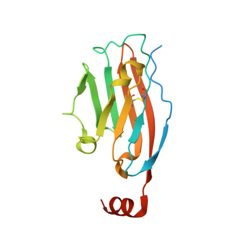
    8P1O

  • PubMed Abstract: 

    Although heavily studied, the subject of anti-PD-L1 small-molecule inhibitors is still elusive. Here we present a systematic overview of the principles behind successful anti-PD-L1 small-molecule inhibitor design on the example of the m -terphenyl scaffold, with a particular focus on the neglected influence of the solubilizer tag on the overall affinity toward PD-L1. The inhibitor developed according to the proposed guidelines was characterized through its potency in blocking PD-1/PD-L1 complex formation in homogeneous time-resolved fluorescence and cell-based assays. The affinity is also explained based on the crystal structure of the inhibitor itself and its costructure with PD-L1 as well as a molecular modeling study. Our results structuralize the knowledge related to the strong pharmacophore feature of the m -terphenyl scaffold preferential geometry and the more complex role of the solubilizer tag in PD-L1 homodimer stabilization.


  • Organizational Affiliation
    • Faculty of Chemistry, Jagiellonian University, Gronostajowa 2, 30-387 Cracow, Poland.

Macromolecule Content 

  • Total Structure Weight: 34.25 kDa 
  • Atom Count: 2,119 
  • Modeled Residue Count: 252 
  • Deposited Residue Count: 288 
  • Unique protein chains: 1

Macromolecules

Find similar proteins by:|  3D Structure
Entity ID: 1
MoleculeChains  Sequence LengthOrganismDetailsImage
Programmed cell death 1 ligand 1
A, B
144Homo sapiensMutation(s): 0 
Gene Names: CD274B7H1PDCD1L1PDCD1LG1PDL1
UniProt & NIH Common Fund Data Resources
Find proteins for Q9NZQ7 (Homo sapiens)
Explore Q9NZQ7 
Go to UniProtKB:  Q9NZQ7
PHAROS:  Q9NZQ7
GTEx:  ENSG00000120217 
Entity Groups
Sequence Clusters30% Identity50% Identity70% Identity90% Identity95% Identity100% Identity
UniProt GroupQ9NZQ7
Sequence Annotations
Expand
Reference Sequence

Experimental Data & Validation

Experimental Data

  • Method: X-RAY DIFFRACTION
  • Resolution: 2.17 Å
  • R-Value Free:  0.289 (Depositor), 0.289 (DCC) 
  • R-Value Work:  0.271 (Depositor), 0.271 (DCC) 
Space Group: P 21 21 2
Unit Cell:
Length ( Å )Angle ( ˚ )
a = 52.088α = 90
b = 109.991β = 90
c = 51.823γ = 90
Software Package:
Software NamePurpose
REFMACrefinement
REFMACrefinement
Aimlessdata scaling
XDSdata reduction
PHASERphasing

Structure Validation

View Full Validation Report


Ligand Structure Quality Assessment 


Entry History 

& Funding Information

Deposition Data


Funding OrganizationLocationGrant Number
Polish National Science CentrePolandUMO-2020/39/D/ST4/01344

Revision History  (Full details and data files)

  • Version 1.0: 2024-01-31
    Type: Initial release
  • Version 1.1: 2024-10-09
    Changes: Structure summary
  • Version 1.2: 2025-12-03
    Changes: Advisory