Detection and characterisation of ligand-induced conformational changes in acetylcholine binding proteins using biosensors and X-ray crystallography.
FitzGerald, E.A., Cederfelt, D., Kovryzhenko, D., Boronat, P., Lund, B.A., Dobritzsch, D., Hennig, S., Paseiro, P.P., de Esch, I.J.P., Danielson, U.H.(2025) RSC Chem Biol 6: 1625-1639
- PubMed: 40896114 Search on PubMedSearch on PubMed Central
- DOI: https://doi.org/10.1039/d5cb00041f
- Primary Citation Related Structures:
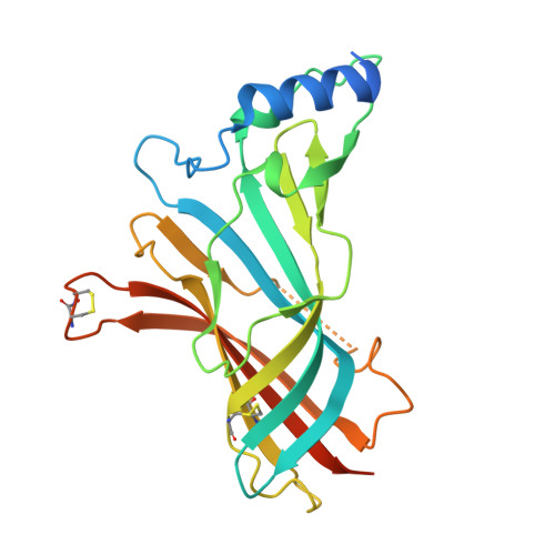
8P11, 8P1E, 8P1F, 8P22, 9SG3 - PubMed Abstract:
Analysis of ligand-induced structural changes in proteins is challenging due to the lack of experimental methods suited for detection and characterisation of both ligand binding and induced structural changes. We have explored biosensors with different detection principles to study interactions between ligands and acetylcholine binding proteins (AChBPs), soluble homologues of Cys-loop ligand gated ion channels (LGICs) that undergo similar structural changes as LGICs upon ligand binding. X-ray crystallography was used to identify binding sites and establish if the detected conformational changes involved small changes in loop C or major structural changes in the pentamer associated with ion channel opening. Experiments were initially focused on ligands exhibiting complex surface plasmon resonance (SPR) biosensor sensorgrams or detected by second harmonic generation (SHG) biosensor analysis. Surface acoustic wave (SAW) and SHG biosensors confirmed that complexities in SPR data were indeed due to ligand-induced conformational changes. Grating coupled interferometry (GCI) biosensor sensorgrams were less complex, despite similar detection principles. switchSENSE biosensor analysis revealed that ligands resulted in either a compaction or expansion of the protein structure. X-ray crystallography of the protein-ligand complexes was only successful for 7 out of 12 ligands, despite nM-μM affinities. Crystals were not obtained for the two compounds shown by SHG analysis to induce large structural changes, while electron densities were not seen in the structures for some ligands. The work presented herein shows that several biosensor technologies have a unique capability to detect and discriminate binding and ligand induced conformational changes in proteins, also when interactions are rapid, weak and structural changes are small. However, they are complementary and provide different information.
- Department of Chemistry - BMC, Uppsala University Sweden helena.danielson@kemi.uu.se.
Organizational Affiliation: