8P1D | pdb_00008p1d

Lysozyme structure solved from serial crystallography data collected at 100 Hz with JUNGFRAU detector at MAXIV


Experimental Data Snapshot

  • Method: X-RAY DIFFRACTION
  • Resolution: 1.60 Å
  • R-Value Free: 
    0.190 (Depositor), 0.190 (DCC) 
  • R-Value Work: 
    0.165 (Depositor), 0.165 (DCC) 
  • R-Value Observed: 
    0.167 (Depositor) 

Starting Model: experimental
View more details

wwPDB Validation 3D Report Full Report

Validation slider image for 8P1D

This is version 1.3 of the entry. See complete history

Literature

Kilohertz serial crystallography with the JUNGFRAU detector at a fourth-generation synchrotron source.

Leonarski, F.Nan, J.Matej, Z.Bertrand, Q.Furrer, A.Gorgisyan, I.Bjelcic, M.Kepa, M.Glover, H.Hinger, V.Eriksson, T.Cehovin, A.Eguiraun, M.Gasparotto, P.Mozzanica, A.Weinert, T.Gonzalez, A.Standfuss, J.Wang, M.Ursby, T.Dworkowski, F.

(2023) IUCrJ 10: 729-737

  • DOI: https://doi.org/10.1107/S2052252523008618
  • Primary Citation Related Structures: 
    8P1A, 8P1B, 8P1C, 8P1D

  • PubMed Abstract: 

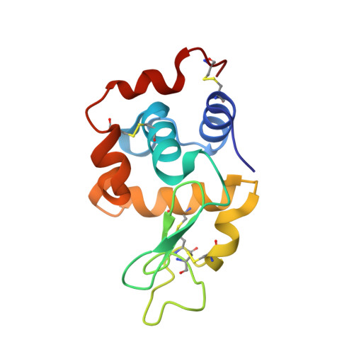
    Serial and time-resolved macromolecular crystallography are on the rise. However, beam time at X-ray free-electron lasers is limited and most third-generation synchrotron-based macromolecular crystallography beamlines do not offer the necessary infrastructure yet. Here, a new setup is demonstrated, based on the JUNGFRAU detector and Jungfraujoch data-acquisition system, that enables collection of kilohertz serial crystallography data at fourth-generation synchrotrons. More importantly, it is shown that this setup is capable of collecting multiple-time-point time-resolved protein dynamics at kilohertz rates, allowing the probing of microsecond to second dynamics at synchrotrons in a fraction of the time needed previously. A high-quality complete X-ray dataset was obtained within 1 min from lysozyme microcrystals, and the dynamics of the light-driven sodium-pump membrane protein KR2 with a time resolution of 1 ms could be demonstrated. To make the setup more accessible for researchers, downstream data handling and analysis will be automated to allow on-the-fly spot finding and indexing, as well as data processing.


  • Organizational Affiliation
    • Photon Science Division, Paul Scherrer Institut, CH-5303 Villigen PSI, Switzerland.

Macromolecule Content 

  • Total Structure Weight: 14.55 kDa 
  • Atom Count: 1,146 
  • Modeled Residue Count: 129 
  • Deposited Residue Count: 129 
  • Unique protein chains: 1

Macromolecules

Find similar proteins by:|  3D Structure
Entity ID: 1
MoleculeChains  Sequence LengthOrganismDetailsImage
Lysozyme C129Gallus gallusMutation(s): 0 
EC: 3.2.1.17
UniProt
Find proteins for P00698 (Gallus gallus)
Explore P00698 
Go to UniProtKB:  P00698
Entity Groups
Sequence Clusters30% Identity50% Identity70% Identity90% Identity95% Identity100% Identity
UniProt GroupP00698
Sequence Annotations
Expand
Reference Sequence

Experimental Data & Validation

Experimental Data

  • Method: X-RAY DIFFRACTION
  • Resolution: 1.60 Å
  • R-Value Free:  0.190 (Depositor), 0.190 (DCC) 
  • R-Value Work:  0.165 (Depositor), 0.165 (DCC) 
  • R-Value Observed: 0.167 (Depositor) 
Space Group: P 43 21 2
Unit Cell:
Length ( Å )Angle ( ˚ )
a = 79.06α = 90
b = 79.06β = 90
c = 38.26γ = 90
Software Package:
Software NamePurpose
PHENIXrefinement
CrystFELdata reduction
CrystFELdata scaling
PHASERphasing

Structure Validation

View Full Validation Report


Entry History 

& Funding Information

Deposition Data


Funding OrganizationLocationGrant Number
Not funded--

Revision History  (Full details and data files)

  • Version 1.0: 2023-10-18
    Type: Initial release
  • Version 1.1: 2023-10-25
    Changes: Database references
  • Version 1.2: 2023-11-15
    Changes: Database references
  • Version 1.3: 2024-11-13
    Changes: Structure summary