8OXX | pdb_00008oxx

Transglutaminase 3 in complex with inhibitor Z-don and DH patient-derived Fab DH63-B02


Experimental Data Snapshot

  • Method: X-RAY DIFFRACTION
  • Resolution: 2.50 Å
  • R-Value Free: 
    0.222 (Depositor), 0.222 (DCC) 
  • R-Value Work: 
    0.154 (Depositor), 0.154 (DCC) 

Starting Model: experimental
View more details

wwPDB Validation 3D Report Full Report

Validation slider image for 8OXX

Ligand Structure Quality Assessment 


This is version 2.0 of the entry. See complete history

Literature

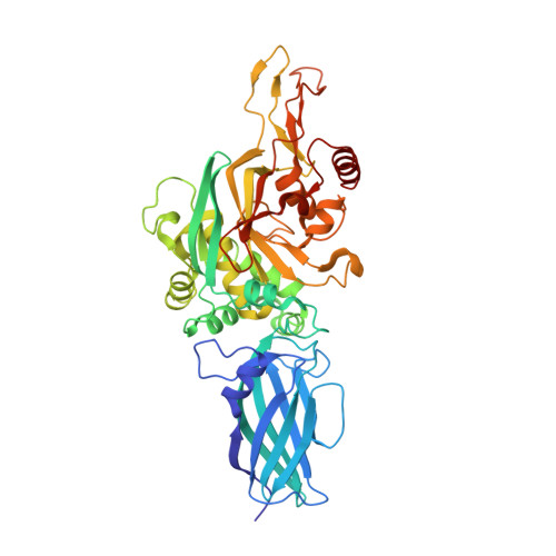
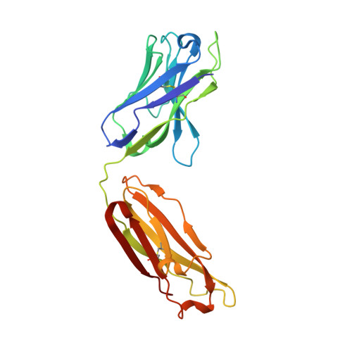
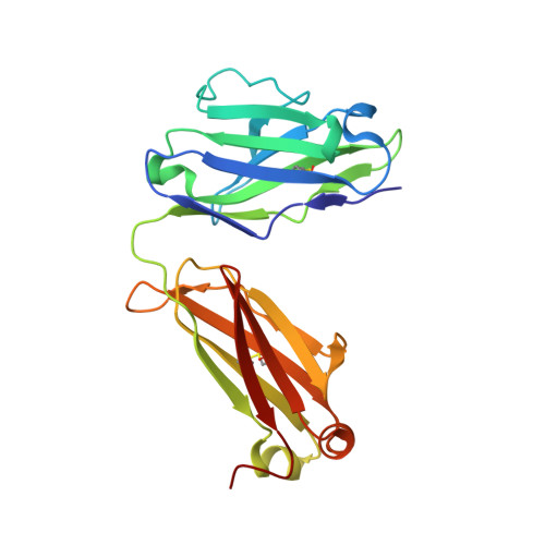
Autoantibody binding and unique enzyme-substrate intermediate conformation of human transglutaminase 3.

Heggelund, J.E.Das, S.Stamnaes, J.Iversen, R.Sollid, L.M.

(2023) Nat Commun 14: 6216-6216

  • DOI: https://doi.org/10.1038/s41467-023-42004-z
  • Primary Citation Related Structures: 
    8OXV, 8OXW, 8OXX, 8OXY

  • PubMed Abstract: 

    Transglutaminase 3 (TG3), the autoantigen of dermatitis herpetiformis (DH), is a calcium dependent enzyme that targets glutamine residues in polypeptides for either transamidation or deamidation modifications. To become catalytically active TG3 requires proteolytic cleavage between the core domain and two C-terminal β-barrels (C1C2). Here, we report four X-ray crystal structures representing inactive and active conformations of human TG3 in complex with a TG3-specific Fab fragment of a DH patient derived antibody. We demonstrate that cleaved TG3, upon binding of a substrate-mimicking inhibitor, undergoes a large conformational change as a β-sheet in the catalytic core domain moves and C1C2 detaches. The unique enzyme-substrate conformation of TG3 without C1C2 is recognized by DH autoantibodies. The findings support a model where B-cell receptors of TG3-specific B cells bind and internalize TG3-gluten enzyme-substrate complexes thereby facilitating gluten-antigen presentation, T-cell help and autoantibody production.


  • Organizational Affiliation
    • KG Jebsen Coeliac Disease Research Centre, Institute of Clinical Medicine, University of Oslo, Oslo, Norway. j.e.heggelund@medisin.uio.no.

Macromolecule Content 

  • Total Structure Weight: 99.26 kDa 
  • Atom Count: 7,433 
  • Modeled Residue Count: 899 
  • Deposited Residue Count: 899 
  • Unique protein chains: 3

Macromolecules

Find similar proteins by:|  3D Structure
Entity ID: 1
MoleculeChains  Sequence LengthOrganismDetailsImage
Protein-glutamine gamma-glutamyltransferase E 27 kDa non-catalytic chain460Homo sapiensMutation(s): 0 
Gene Names: TGM3
EC: 2.3.2.13
UniProt & NIH Common Fund Data Resources
Find proteins for Q08188 (Homo sapiens)
Explore Q08188 
Go to UniProtKB:  Q08188
PHAROS:  Q08188
GTEx:  ENSG00000125780 
Entity Groups
Sequence Clusters30% Identity50% Identity70% Identity90% Identity95% Identity100% Identity
UniProt GroupQ08188
Sequence Annotations
Expand
Reference Sequence
Find similar proteins by:|  3D Structure
Entity ID: 2
MoleculeChains  Sequence LengthOrganismDetailsImage
Antibody fab fragment heavy chain223Homo sapiensMutation(s): 0 
Find similar proteins by:|  3D Structure
Entity ID: 3
MoleculeChains  Sequence LengthOrganismDetailsImage
Antibody fab fragment light chain216Homo sapiensMutation(s): 0 

Small Molecules

Ligands 8 Unique
IDChains Name / Formula / InChI Key2D Diagram3D Interactions
P6S
(Subject of Investigation/LOI)

Query on P6S


Download:Ideal Coordinates CCD File
J [auth A]benzyl hydrogen carbonate
C8 H8 O3
NJAPCAIWQRPQPY-UHFFFAOYSA-N
MLL
(Subject of Investigation/LOI)

Query on MLL


Download:Ideal Coordinates CCD File
N [auth A]METHYL L-LEUCINATE
C7 H15 N O2
QVDXUKJJGUSGLS-LURJTMIESA-N
ONL
(Subject of Investigation/LOI)

Query on ONL


Download:Ideal Coordinates CCD File
K [auth A]5-OXO-L-NORLEUCINE
C6 H11 N O3
KSIJECNNZVKMJG-YFKPBYRVSA-N
DVA
(Subject of Investigation/LOI)

Query on DVA


Download:Ideal Coordinates CCD File
L [auth A]D-VALINE
C5 H11 N O2
KZSNJWFQEVHDMF-SCSAIBSYSA-N
DPR
(Subject of Investigation/LOI)

Query on DPR


Download:Ideal Coordinates CCD File
M [auth A]D-PROLINE
C5 H9 N O2
ONIBWKKTOPOVIA-SCSAIBSYSA-N
SO4

Query on SO4


Download:Ideal Coordinates CCD File
D [auth A]SULFATE ION
O4 S
QAOWNCQODCNURD-UHFFFAOYSA-L
EDO

Query on EDO


Download:Ideal Coordinates CCD File
E [auth A]
F [auth A]
O [auth B]
P [auth B]
Q [auth C]
E [auth A],
F [auth A],
O [auth B],
P [auth B],
Q [auth C],
R [auth C]
1,2-ETHANEDIOL
C2 H6 O2
LYCAIKOWRPUZTN-UHFFFAOYSA-N
CA

Query on CA


Download:Ideal Coordinates CCD File
G [auth A],
H [auth A],
I [auth A]
CALCIUM ION
Ca
BHPQYMZQTOCNFJ-UHFFFAOYSA-N

Experimental Data & Validation

Experimental Data

  • Method: X-RAY DIFFRACTION
  • Resolution: 2.50 Å
  • R-Value Free:  0.222 (Depositor), 0.222 (DCC) 
  • R-Value Work:  0.154 (Depositor), 0.154 (DCC) 
Space Group: P 1 21 1
Unit Cell:
Length ( Å )Angle ( ˚ )
a = 79.271α = 90
b = 65.005β = 96.854
c = 90.908γ = 90
Software Package:
Software NamePurpose
REFMACrefinement
REFMACrefinement
Cootmodel building
PHASERphasing
XDSdata reduction
Aimlessdata scaling

Structure Validation

View Full Validation Report


Ligand Structure Quality Assessment 


Entry History 

& Funding Information

Deposition Data


Funding OrganizationLocationGrant Number
Other governmentNorway2020027
Other privateNorwaySKGJ-MED-017
Other governmentNorwayWL-IMMUNOLOGY

Revision History  (Full details and data files)

  • Version 1.0: 2023-10-18
    Type: Initial release
  • Version 2.0: 2023-11-15
    Changes: Atomic model, Data collection