Exploring the pH dependence of an improved PETase.
Charlier, C., Gavalda, S., Grga, J., Perrot, L., Gabrielli, V., Lohr, F., Schorghuber, J., Lichtenecker, R., Arnal, G., Marty, A., Tournier, V., Lippens, G.(2024) Biophys J 123: 1542-1552
- PubMed: 38664965 Search on PubMedSearch on PubMed Central
- DOI: https://doi.org/10.1016/j.bpj.2024.04.026
- Primary Citation Related Structures:
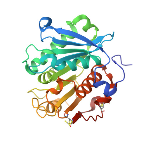
8OTA - PubMed Abstract:
Enzymatic recycling of plastic and especially of polyethylene terephthalate (PET) has shown great potential to reduce its negative impact on our society. PET hydrolases (PETases) have been optimized using rational design and machine learning, but the mechanistic details of the PET depolymerization process remain unclear. Belonging to the carboxylic-ester hydrolase family with a canonical Ser-His-Asp catalytic triad, their observed alkaline pH optimum is generally thought to be related to the protonation state of the catalytic His. Here, we explore this aspect in the context of LCC ICCG , an optimized PETase, derived from the leaf-branch compost cutinase enzyme. We use NMR to identify the dominant tautomeric structure of the six histidines. Five show surprisingly low pKa values below 4.0, whereas the catalytic H242 in the active enzyme displays a pKa value that varies from 4.9 to 4.7 when temperatures increase from 30°C to 50°C. Whereas the hydrolytic activity of the enzyme toward a soluble substrate can be modeled by the corresponding protonation/deprotonation curve, an important discrepancy is found when the substrate is the solid plastic. This opens the way to further mechanistic understanding of the PETase activity and underscores the importance of studying the enzyme at the liquid-solid interface.
- Toulouse Biotechnology Institute (TBI), University of Toulouse, CNRS, INRAE, INSA Toulouse, Toulouse Cedex, France.
Organizational Affiliation: