8OS6 | pdb_00008os6

Structure of a GFRA1/GDNF LICAM complex


Experimental Data Snapshot

  • Method: X-RAY DIFFRACTION
  • Resolution: 2.66 Å
  • R-Value Free: 
    0.283 (Depositor), 0.253 (DCC) 
  • R-Value Work: 
    0.244 (Depositor), 0.245 (DCC) 
  • R-Value Observed: 
    0.246 (Depositor) 

Starting Model: experimental
View more details

wwPDB Validation 3D Report Full Report

Validation slider image for 8OS6

Ligand Structure Quality Assessment 


This is version 1.2 of the entry. See complete history

Literature

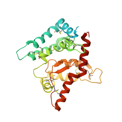
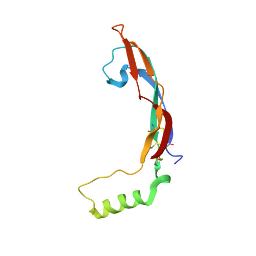
Architecture and regulation of a GDNF-GFR alpha 1 synaptic adhesion assembly.

Houghton, F.M.Adams, S.E.Rios, A.S.Masino, L.Purkiss, A.G.Briggs, D.C.Ledda, F.McDonald, N.Q.

(2023) Nat Commun 14: 7551-7551

  • DOI: https://doi.org/10.1038/s41467-023-43148-8
  • Primary Citation Related Structures: 
    8OS6

  • PubMed Abstract: 

    Glial-cell line derived neurotrophic factor (GDNF) bound to its co-receptor GFRα1 stimulates the RET receptor tyrosine kinase, promoting neuronal survival and neuroprotection. The GDNF-GFRα1 complex also supports synaptic cell adhesion independently of RET. Here, we describe the structure of a decameric GDNF-GFRα1 assembly determined by crystallography and electron microscopy, revealing two GFRα1 pentamers bridged by five GDNF dimers. We reconsitituted the assembly between adhering liposomes and used cryo-electron tomography to visualize how the complex fulfils its membrane adhesion function. The GFRα1:GFRα1 pentameric interface was further validated both in vitro by native PAGE and in cellulo by cell-clustering and dendritic spine assays. Finally, we provide biochemical and cell-based evidence that RET and heparan sulfate cooperate to prevent assembly of the adhesion complex by competing for the adhesion interface. Our results provide a mechanistic framework to understand GDNF-driven cell adhesion, its relationship to trophic signalling, and the central role played by GFRα1.


  • Organizational Affiliation
    • Signalling and Structural Biology laboratory, The Francis Crick Institute, 1 Midland Road, London, NW1 1AT, UK.

Macromolecule Content 

  • Total Structure Weight: 411.87 kDa 
  • Atom Count: 23,825 
  • Modeled Residue Count: 3,001 
  • Deposited Residue Count: 3,590 
  • Unique protein chains: 2

Macromolecules

Find similar proteins by:|  3D Structure
Entity ID: 1
MoleculeChains  Sequence LengthOrganismDetailsImage
GDNF family receptor alpha
A, C, E, G, I
A, C, E, G, I, K, M, O, Q, S
260Danio rerioMutation(s): 0 
Gene Names: gfralpha1azgc:110129gfra1a
UniProt
Find proteins for Q98TT9 (Danio rerio)
Explore Q98TT9 
Go to UniProtKB:  Q98TT9
Entity Groups
Sequence Clusters30% Identity50% Identity70% Identity90% Identity95% Identity100% Identity
UniProt GroupQ98TT9
Glycosylation
Glycosylation Sites: 1
Sequence Annotations
Expand
Reference Sequence
Find similar proteins by:|  3D Structure
Entity ID: 2
MoleculeChains  Sequence LengthOrganismDetailsImage
Glial cell line-derived neurotrophic factor
B, D, F, H, J
B, D, F, H, J, L, N, P, R, T
99Danio rerioMutation(s): 0 
Gene Names: gdnf
UniProt
Find proteins for Q98TU0 (Danio rerio)
Explore Q98TU0 
Go to UniProtKB:  Q98TU0
Entity Groups
Sequence Clusters30% Identity50% Identity70% Identity90% Identity95% Identity100% Identity
UniProt GroupQ98TU0
Glycosylation
Glycosylation Sites: 1
Sequence Annotations
Expand
Reference Sequence

Oligosaccharides

Help  
Entity ID: 3
MoleculeChains Length2D Diagram GlycosylationD Interactions
2-acetamido-2-deoxy-beta-D-glucopyranose-(1-4)-2-acetamido-2-deoxy-beta-D-glucopyranoseBA [auth b],
U,
Y,
Z
2N-Glycosylation
Glycosylation Resources
GlyTouCan: G42666HT
GlyCosmos: G42666HT
GlyGen: G42666HT
Entity ID: 4
MoleculeChains Length2D Diagram GlycosylationD Interactions
2-acetamido-2-deoxy-beta-D-glucopyranose-(1-4)-[alpha-L-fucopyranose-(1-6)]2-acetamido-2-deoxy-beta-D-glucopyranose
V, W, X
3N-Glycosylation
Glycosylation Resources
GlyTouCan: G21290RB
GlyCosmos: G21290RB
GlyGen: G21290RB
Entity ID: 5
MoleculeChains Length2D Diagram GlycosylationD Interactions
alpha-L-fucopyranose-(1-6)-2-acetamido-2-deoxy-beta-D-glucopyranoseAA [auth a]2N-Glycosylation
Glycosylation Resources
GlyTouCan: G86851RC
GlyCosmos: G86851RC
GlyGen: G86851RC

Small Molecules

Ligands 1 Unique
IDChains Name / Formula / InChI Key2D Diagram3D Interactions
NAG

Query on NAG


Download:Ideal Coordinates CCD File
CA [auth B]
DA [auth E]
EA [auth G]
FA [auth I]
GA [auth K]
CA [auth B],
DA [auth E],
EA [auth G],
FA [auth I],
GA [auth K],
HA [auth L],
IA [auth M],
JA [auth R],
KA [auth S],
LA [auth T]
2-acetamido-2-deoxy-beta-D-glucopyranose
C8 H15 N O6
OVRNDRQMDRJTHS-FMDGEEDCSA-N

Experimental Data & Validation

Experimental Data

  • Method: X-RAY DIFFRACTION
  • Resolution: 2.66 Å
  • R-Value Free:  0.283 (Depositor), 0.253 (DCC) 
  • R-Value Work:  0.244 (Depositor), 0.245 (DCC) 
  • R-Value Observed: 0.246 (Depositor) 
Space Group: P 1 21 1
Unit Cell:
Length ( Å )Angle ( ˚ )
a = 114.14α = 90
b = 170.044β = 96.17
c = 130.791γ = 90
Software Package:
Software NamePurpose
PHENIXrefinement
DIALSdata reduction
DIALSdata scaling
PHASERphasing

Structure Validation

View Full Validation Report


Ligand Structure Quality Assessment 


Entry History 

& Funding Information

Deposition Data


Funding OrganizationLocationGrant Number
The Francis Crick InstituteUnited KingdomCC2068

Revision History  (Full details and data files)

  • Version 1.0: 2023-11-29
    Type: Initial release
  • Version 1.1: 2024-03-27
    Changes: Database references
  • Version 1.2: 2024-11-20
    Changes: Structure summary