8ORM | pdb_00008orm

Cryo-EM structure of CAK-THZ1


Experimental Data Snapshot

  • Method: ELECTRON MICROSCOPY
  • Resolution: 1.90 Å
  • Aggregation State: PARTICLE 
  • Reconstruction Method: SINGLE PARTICLE 

Starting Model: experimental
View more details

wwPDB Validation 3D Report Full Report

Validation slider image for 8ORM

This is version 1.3 of the entry. See complete history. 

Literature

High-resolution cryo-EM of the human CDK-activating kinase for structure-based drug design.

Cushing, V.I., Koh, A.F., Feng, J., Jurgaityte, K., Bondke, A., Kroll, S.H.B., Barbazanges, M., Scheiper, B., Bahl, A.K., Barrett, A.G.M., Ali, S., Kotecha, A., Greber, B.J.

(2024) Nat Commun 15: 2265-2265

  • DOI: https://doi.org/10.1038/s41467-024-46375-9
  • Primary Citation Related Structures: 
    8ORM, 8P6V, 8P6W, 8P6X, 8P6Y, 8P6Z, 8P70, 8P71, 8P72, 8P73, 8P74, 8P75, 8P76, 8P77, 8P78, 8P79, 8P7L, 8PLZ

  • PubMed Abstract: 

    Rational design of next-generation therapeutics can be facilitated by high-resolution structures of drug targets bound to small-molecule inhibitors. However, application of structure-based methods to macromolecules refractory to crystallization has been hampered by the often-limiting resolution and throughput of cryogenic electron microscopy (cryo-EM). Here, we use high-resolution cryo-EM to determine structures of the CDK-activating kinase, a master regulator of cell growth and division, in its free and nucleotide-bound states and in complex with 15 inhibitors at up to 1.8 Å resolution. Our structures provide detailed insight into inhibitor interactions and networks of water molecules in the active site of cyclin-dependent kinase 7 and provide insights into the mechanisms contributing to inhibitor selectivity, thereby providing the basis for rational design of next-generation therapeutics. These results establish a methodological framework for the use of high-resolution cryo-EM in structure-based drug design.


  • Organizational Affiliation: 
    • The Institute of Cancer Research, Chester Beatty Laboratories, 237 Fulham Road, London, SW3 6JB, UK.

Macromolecule Content 

  • Total Structure Weight: 87.89 kDa 
  • Atom Count: 5,341 
  • Modeled Residue Count: 633 
  • Deposited Residue Count: 766 
  • Unique protein chains: 3

Macromolecules

Find similar proteins by:|  3D Structure
Entity ID: 1
MoleculeChains  Sequence LengthOrganismDetailsImage
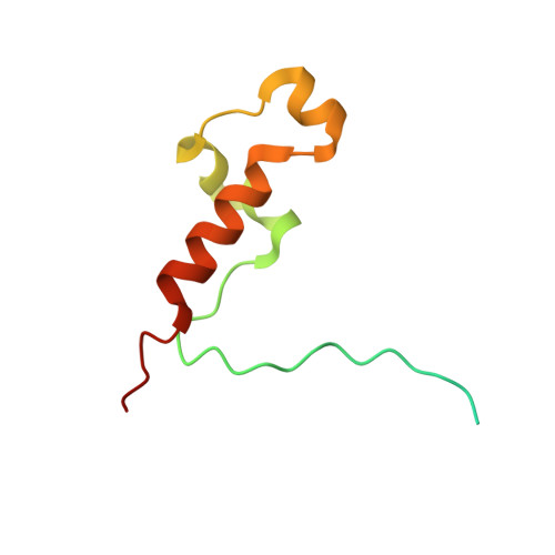
CDK-activating kinase assembly factor MAT1A [auth H]93Homo sapiensMutation(s): 0 
Gene Names: MNAT1, CAP35, MAT1, RNF66
UniProt & NIH Common Fund Data Resources
Find proteins for P51948 (Homo sapiens)
Explore P51948 
Go to UniProtKB:  P51948
PHAROS:  P51948
GTEx:  ENSG00000020426 
Entity Groups
Sequence Clusters30% Identity50% Identity70% Identity90% Identity95% Identity100% Identity
UniProt GroupP51948
Sequence Annotations
Expand
Reference Sequence
Find similar proteins by:|  3D Structure
Entity ID: 2
MoleculeChains  Sequence LengthOrganismDetailsImage
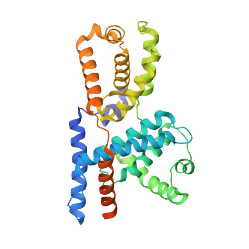
Cyclin-HB [auth I]324Homo sapiensMutation(s): 1 
Gene Names: CCNH
UniProt & NIH Common Fund Data Resources
Find proteins for P51946 (Homo sapiens)
Explore P51946 
Go to UniProtKB:  P51946
PHAROS:  P51946
GTEx:  ENSG00000134480 
Entity Groups
Sequence Clusters30% Identity50% Identity70% Identity90% Identity95% Identity100% Identity
UniProt GroupP51946
Sequence Annotations
Expand
Reference Sequence
Find similar proteins by:|  3D Structure
Entity ID: 3
MoleculeChains  Sequence LengthOrganismDetailsImage
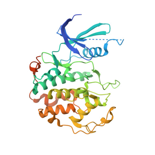
Cyclin-dependent kinase 7C [auth J]349Homo sapiensMutation(s): 0 
Gene Names: CDK7, CAK, CAK1, CDKN7, MO15, STK1
EC: 2.7.11.22 (PDB Primary Data), 2.7.11.23 (PDB Primary Data)
UniProt & NIH Common Fund Data Resources
Find proteins for P50613 (Homo sapiens)
Explore P50613 
Go to UniProtKB:  P50613
PHAROS:  P50613
GTEx:  ENSG00000134058 
Entity Groups
Sequence Clusters30% Identity50% Identity70% Identity90% Identity95% Identity100% Identity
UniProt GroupP50613
Sequence Annotations
Expand
Reference Sequence

Small Molecules

Ligands 1 Unique
IDChains Name / Formula / InChI Key2D Diagram3D Interactions
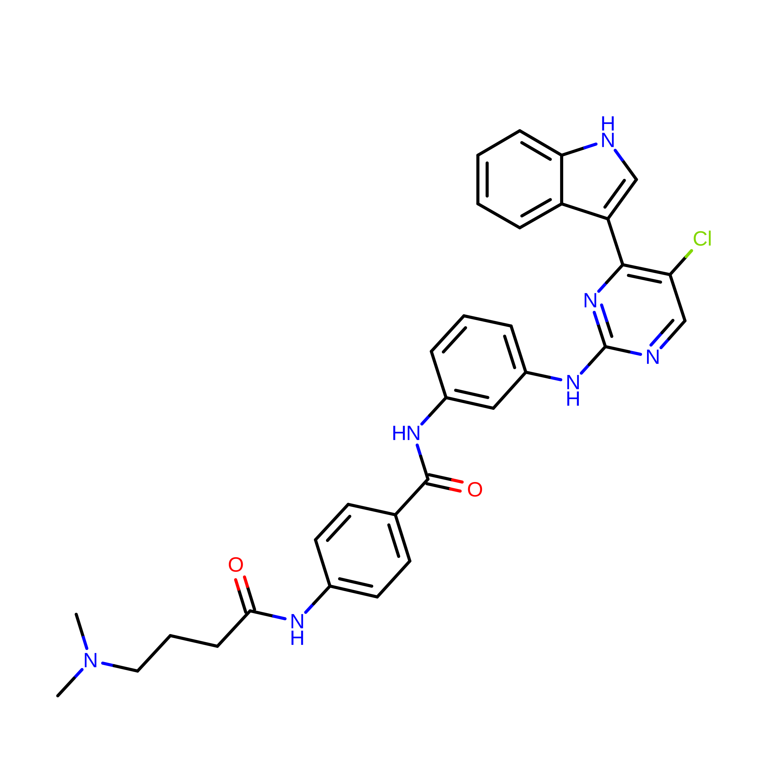
V0G
(Subject of Investigation/LOI)

Query on V0G


Download:Ideal Coordinates CCD File
D [auth J]N-(3-{[5-chloro-4-(1H-indol-3-yl)pyrimidin-2-yl]amino}phenyl)-4-{[4-(dimethylamino)butanoyl]amino}benzamide
C31 H30 Cl N7 O2
TUERFPPIPKZNKE-UHFFFAOYSA-N

Experimental Data & Validation

Experimental Data

  • Method: ELECTRON MICROSCOPY
  • Resolution: 1.90 Å
  • Aggregation State: PARTICLE 
  • Reconstruction Method: SINGLE PARTICLE 
EM Software:
TaskSoftware PackageVersion
RECONSTRUCTIONRELION4.0 beta
MODEL REFINEMENTPHENIX1.20.1

Structure Validation

View Full Validation Report


Entry History 

& Funding Information

Deposition Data


Funding OrganizationLocationGrant Number
Medical Research Council (MRC, United Kingdom)United KingdomMR/V009354/1

Revision History  (Full details and data files)

  • Version 1.0: 2023-08-30
    Type: Initial release
  • Version 1.1: 2024-03-20
    Changes: Database references, Refinement description
  • Version 1.2: 2024-03-27
    Changes: Database references
  • Version 1.3: 2024-11-13
    Changes: Data collection, Structure summary