8OR7 | pdb_00008or7

Structure of a far-red induced allophycocyanin from Chroococcidiopsis thermalis sp. PCC 7203


Experimental Data Snapshot

  • Method: X-RAY DIFFRACTION
  • Resolution: 2.80 Å
  • R-Value Free: 
    0.240 (Depositor), 0.238 (DCC) 
  • R-Value Work: 
    0.178 (Depositor), 0.182 (DCC) 
  • R-Value Observed: 
    0.181 (Depositor) 

Starting Models: experimental
View more details

wwPDB Validation 3D Report Full Report

Validation slider image for 8OR7

Ligand Structure Quality Assessment 


This is version 1.2 of the entry. See complete history

Literature

Crystallographic and biochemical analyses of a far-red allophycocyanin to address the mechanism of the super-red-shift.

Zhou, L.J.Hoppner, A.Wang, Y.Q.Hou, J.Y.Scheer, H.Zhao, K.H.

(2024) Photosynth Res 162: 171-185

  • DOI: https://doi.org/10.1007/s11120-023-01066-2
  • Primary Citation Related Structures: 
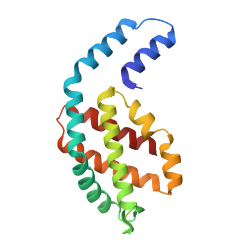
    8OR7

  • PubMed Abstract: 

    Far-red absorbing allophycocyanins (APC), identified in cyanobacteria capable of FRL photoacclimation (FaRLiP) and low-light photoacclimation (LoLiP), absorb far-red light, functioning in energy transfer as light-harvesting proteins. We report an optimized method to obtain high purity far-red absorbing allophycocyanin B, AP-B2, of Chroococcidiopsis thermalis sp. PCC7203 by synthesis in Escherichia coli and an improved purification protocol. The crystal structure of the trimer, (PCB-ApcD5/PCB-ApcB2) 3 , has been resolved to 2.8 Å. The main difference to conventional APCs absorbing in the 650-670 nm range is a largely flat chromophore with the co-planarity extending, in particular, from rings BCD to ring A. This effectively extends the conjugation system of PCB and contributes to the super-red-shifted absorption of the α-subunit (λ max  = 697 nm). On complexation with the β-subunit, it is even further red-shifted (λ max, absorption  = 707 nm, λ max, emission  = 721 nm). The relevance of ring A for this shift is supported by mutagenesis data. A variant of the α-subunit, I123M, has been generated that shows an intense FR-band already in the absence of the β-subunit, a possible model is discussed. Two additional mechanisms are known to red-shift the chromophore spectrum: lactam-lactim tautomerism and deprotonation of the chromophore that both mechanisms appear inconsistent with our data, leaving this question unresolved.


  • Organizational Affiliation
    • National Key Laboratory of Agricultural Microbiology, Huazhong Agricultural University, Wuhan, 430070, The People's Republic of China.

Macromolecule Content 

  • Total Structure Weight: 76.25 kDa 
  • Atom Count: 5,179 
  • Modeled Residue Count: 640 
  • Deposited Residue Count: 656 
  • Unique protein chains: 2

Macromolecules

Find similar proteins by:|  3D Structure
Entity ID: 1
MoleculeChains  Sequence LengthOrganismDetailsImage
Allophycocyanin beta subunit apoproteinA [auth B],
B [auth D]
169Chroococcidiopsis thermalisMutation(s): 0 
Gene Names: Chro_1033
UniProt
Find proteins for K9TVG8 (Chroococcidiopsis thermalis (strain PCC 7203))
Explore K9TVG8 
Go to UniProtKB:  K9TVG8
Entity Groups
Sequence Clusters30% Identity50% Identity70% Identity90% Identity95% Identity100% Identity
UniProt GroupK9TVG8
Sequence Annotations
Expand
Reference Sequence
Find similar proteins by:|  3D Structure
Entity ID: 2
MoleculeChains  Sequence LengthOrganismDetailsImage
PhycocyaninC [auth A],
D [auth C]
159Chroococcidiopsis thermalisMutation(s): 0 
Gene Names: Chro_1032
UniProt
Find proteins for K9TX42 (Chroococcidiopsis thermalis (strain PCC 7203))
Explore K9TX42 
Go to UniProtKB:  K9TX42
Entity Groups
Sequence Clusters30% Identity50% Identity70% Identity90% Identity95% Identity100% Identity
UniProt GroupK9TX42
Sequence Annotations
Expand
Reference Sequence

Small Molecules

Ligands 3 Unique
IDChains Name / Formula / InChI Key2D Diagram3D Interactions
CYC
(Subject of Investigation/LOI)

Query on CYC


Download:Ideal Coordinates CCD File
E [auth B],
H [auth D],
I [auth A],
L [auth C]
PHYCOCYANOBILIN
C33 H40 N4 O6
VXTXPYZGDQPMHK-GMXXPEQVSA-N
SO4

Query on SO4


Download:Ideal Coordinates CCD File
G [auth B],
M [auth C],
O [auth C]
SULFATE ION
O4 S
QAOWNCQODCNURD-UHFFFAOYSA-L
K

Query on K


Download:Ideal Coordinates CCD File
F [auth B],
J [auth A],
K [auth A],
N [auth C]
POTASSIUM ION
K
NPYPAHLBTDXSSS-UHFFFAOYSA-N

Experimental Data & Validation

Experimental Data

  • Method: X-RAY DIFFRACTION
  • Resolution: 2.80 Å
  • R-Value Free:  0.240 (Depositor), 0.238 (DCC) 
  • R-Value Work:  0.178 (Depositor), 0.182 (DCC) 
  • R-Value Observed: 0.181 (Depositor) 
Space Group: P 63
Unit Cell:
Length ( Å )Angle ( ˚ )
a = 130.54α = 90
b = 130.54β = 90
c = 106.496γ = 120
Software Package:
Software NamePurpose
REFMACrefinement
xia2data reduction
pointlessdata scaling
ARP/wARPphasing

Structure Validation

View Full Validation Report


Ligand Structure Quality Assessment 


Entry History 

& Funding Information

Deposition Data


Funding OrganizationLocationGrant Number
National Natural Science Foundation of China (NSFC)China31861143029

Revision History  (Full details and data files)

  • Version 1.0: 2024-03-20
    Type: Initial release
  • Version 1.1: 2024-11-06
    Changes: Structure summary
  • Version 1.2: 2024-12-11
    Changes: Database references