8OO9 | pdb_00008oo9

CryoEM Structure INO80core Hexasome complex ATPase-DNA refinement state1


Experimental Data Snapshot

  • Method: ELECTRON MICROSCOPY
  • Resolution: 3.20 Å
  • Aggregation State: PARTICLE 
  • Reconstruction Method: SINGLE PARTICLE 

Starting Models: experimental
View more details

wwPDB Validation 3D Report Full Report

Validation slider image for 8OO9

This is version 1.2 of the entry. See complete history

Re-refinement Note

A newer entry is available that reflects an alternative modeling of the original data: 8OO9

Literature

Hexasome-INO80 complex reveals structural basis of noncanonical nucleosome remodeling.

Zhang, M.Jungblut, A.Kunert, F.Hauptmann, L.Hoffmann, T.Kolesnikova, O.Metzner, F.Moldt, M.Weis, F.DiMaio, F.Hopfner, K.P.Eustermann, S.

(2023) Science 381: 313-319

  • DOI: https://doi.org/10.1126/science.adf6287
  • Primary Citation Related Structures: 
    8OO7, 8OO9, 8OOA, 8OOC, 8OOF, 8OOK, 8OOP, 8OOR, 8OOS, 8OOT

  • PubMed Abstract: 

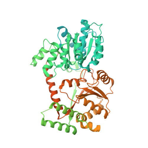
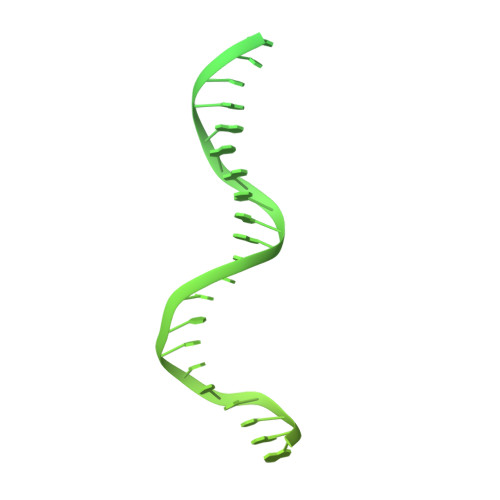
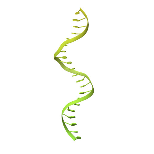
    Loss of H2A-H2B histone dimers is a hallmark of actively transcribed genes, but how the cellular machinery functions in the context of noncanonical nucleosomal particles remains largely elusive. In this work, we report the structural mechanism for adenosine 5'-triphosphate-dependent chromatin remodeling of hexasomes by the INO80 complex. We show how INO80 recognizes noncanonical DNA and histone features of hexasomes that emerge from the loss of H2A-H2B. A large structural rearrangement switches the catalytic core of INO80 into a distinct, spin-rotated mode of remodeling while its nuclear actin module remains tethered to long stretches of unwrapped linker DNA. Direct sensing of an exposed H3-H4 histone interface activates INO80, independently of the H2A-H2B acidic patch. Our findings reveal how the loss of H2A-H2B grants remodelers access to a different, yet unexplored layer of energy-driven chromatin regulation.


  • Organizational Affiliation
    • Structural and Computational Biology Unit, European Molecular Biology Laboratory (EMBL), Heidelberg, Germany.

Macromolecule Content 

  • Total Structure Weight: 271.01 kDa 
  • Atom Count: 4,516 
  • Modeled Residue Count: 481 
  • Deposited Residue Count: 1,586 
  • Unique protein chains: 1
  • Unique nucleic acid chains: 2

Macromolecules


Find similar proteins by:|  3D Structure
Entity ID: 1
MoleculeChains  Sequence LengthOrganismDetailsImage
Chromatin-remodeling ATPase INO80A [auth G]1,134Thermochaetoides thermophilaMutation(s): 0 
Gene Names: TT172_LOCUS3790
Find similar nucleic acids by:  (by identity cutoff) 
Entity ID: 2
MoleculeChains LengthOrganismImage
DNA strand 1B [auth K]226synthetic construct
Sequence Annotations
Expand
Reference Sequence
Find similar nucleic acids by:  (by identity cutoff) 
Entity ID: 3
MoleculeChains LengthOrganismImage
DNA strand 2C [auth L]226synthetic construct
Sequence Annotations
Expand
Reference Sequence

Experimental Data & Validation

Experimental Data

  • Method: ELECTRON MICROSCOPY
  • Resolution: 3.20 Å
  • Aggregation State: PARTICLE 
  • Reconstruction Method: SINGLE PARTICLE 
EM Software:
TaskSoftware PackageVersion
RECONSTRUCTIONRELION4.0
MODEL REFINEMENTPHENIX1.20.1

Structure Validation

View Full Validation Report


Entry History 

& Funding Information

Deposition Data


Funding OrganizationLocationGrant Number
EIPOD fellowship under Marie Sklodowska-Curie Actions COFUNDGermany847543
European Research Council (ERC)European Union833613
German Research Foundation (DFG)GermanyCRC136
German Research Foundation (DFG)GermanyCRC1064

Revision History  (Full details and data files)

  • Version 1.0: 2023-07-26
    Type: Initial release
  • Version 1.1: 2023-08-02
    Changes: Database references
  • Version 1.2: 2024-07-24
    Changes: Data collection, Refinement description