8ONX | pdb_00008onx

High resolution structure of Chaetomium thermophilum MAP2


Experimental Data Snapshot

  • Method: X-RAY DIFFRACTION
  • Resolution: 1.30 Å
  • R-Value Free: 
    0.177 (Depositor), 0.177 (DCC) 
  • R-Value Work: 
    0.161 (Depositor), 0.161 (DCC) 
  • R-Value Observed: 
    0.162 (Depositor) 

Starting Model: in silico
View more details

wwPDB Validation 3D Report Full Report

Validation slider image for 8ONX

This is version 1.0 of the entry. See complete history

Literature

Methionine aminopeptidase 2 and its autoproteolysis product have different binding sites on the ribosome.

Klein, M.A.Wild, K.Kisonaite, M.Sinning, I.

(2024) Nat Commun 15: 716-716

  • DOI: https://doi.org/10.1038/s41467-024-44862-7
  • Primary Citation Related Structures: 
    8ONX, 8ONY, 8ONZ, 8OO0

  • PubMed Abstract: 

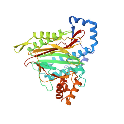
    Excision of the initiator methionine is among the first co-translational processes that occur at the ribosome. While this crucial step in protein maturation is executed by two types of methionine aminopeptidases in eukaryotes (MAP1 and MAP2), additional roles in disease and translational regulation have drawn more attention to MAP2. Here, we report several cryo-EM structures of human and fungal MAP2 at the 80S ribosome. Irrespective of nascent chains, MAP2 can occupy the tunnel exit. On nascent chain displaying ribosomes, the MAP2-80S interaction is highly dynamic and the MAP2-specific N-terminal extension engages in stabilizing interactions with the long rRNA expansion segment ES27L. Loss of this extension by autoproteolytic cleavage impedes interactions at the tunnel, while promoting MAP2 to enter the ribosomal A-site, where it engages with crucial functional centers of translation. These findings reveal that proteolytic remodeling of MAP2 severely affects ribosome binding, and set the stage for targeted functional studies.


  • Organizational Affiliation
    • Heidelberg University Biochemistry Center (BZH), Im Neuenheimer Feld 328, 69120, Heidelberg, Germany.

Macromolecule Content 

  • Total Structure Weight: 41.81 kDa 
  • Atom Count: 3,509 
  • Modeled Residue Count: 371 
  • Deposited Residue Count: 376 
  • Unique protein chains: 1

Macromolecules

Find similar proteins by:|  3D Structure
Entity ID: 1
MoleculeChains  Sequence LengthOrganismDetailsImage
Methionine aminopeptidase 2376Thermochaetoides thermophilaMutation(s): 0 
Gene Names: CTHT_0063100
EC: 3.4.11.18
UniProt
Find proteins for G0SEA9 (Chaetomium thermophilum (strain DSM 1495 / CBS 144.50 / IMI 039719))
Explore G0SEA9 
Go to UniProtKB:  G0SEA9
Entity Groups
Sequence Clusters30% Identity50% Identity70% Identity90% Identity95% Identity100% Identity
UniProt GroupG0SEA9
Sequence Annotations
Expand
Reference Sequence

Experimental Data & Validation

Experimental Data

  • Method: X-RAY DIFFRACTION
  • Resolution: 1.30 Å
  • R-Value Free:  0.177 (Depositor), 0.177 (DCC) 
  • R-Value Work:  0.161 (Depositor), 0.161 (DCC) 
  • R-Value Observed: 0.162 (Depositor) 
Space Group: P 21 21 21
Unit Cell:
Length ( Å )Angle ( ˚ )
a = 59.938α = 90
b = 73.806β = 90
c = 82.049γ = 90
Software Package:
Software NamePurpose
PHENIXrefinement
XDSdata reduction
Aimlessdata scaling
PHENIXphasing

Structure Validation

View Full Validation Report


Entry History 

& Funding Information

Deposition Data


Funding OrganizationLocationGrant Number
German Research Foundation (DFG)GermanySI 586-6

Revision History  (Full details and data files)

  • Version 1.0: 2024-02-14
    Type: Initial release