8OMF

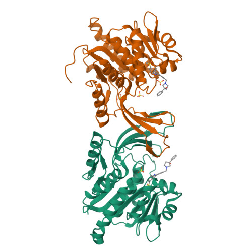
Crystal structure of hKHK-C in complex with compound-4


Experimental Data Snapshot

  • Method: X-RAY DIFFRACTION
  • Resolution: 2.14 Å
  • R-Value Free: 0.254 
  • R-Value Work: 0.228 
  • R-Value Observed: 0.229 

Starting Model: experimental
View more details

wwPDB Validation   3D Report Full Report


Ligand Structure Quality Assessment 


This is version 1.1 of the entry. See complete history


Literature

Crystal structures of human and mouse ketohexokinase provide a structural basis for species- and isoform-selective inhibitor design.

Ebenhoch, R.Bauer, M.Romig, H.Gottschling, D.Kley, J.T.Heine, N.Weber, A.Uphues, I.Nar, H.Pautsch, A.

(2023) Acta Crystallogr D Struct Biol 79: 871-880

  • DOI: https://doi.org/10.1107/S2059798323006137
  • Primary Citation of Related Structures:  
    8OMD, 8OME, 8OMF, 8OMG

  • PubMed Abstract: 

    A molecular understanding of the proteins involved in fructose metabolism is essential for controlling the current spread of fructose-related obesity, diabetes and related adverse metabolic states in Western populations. Fructose catabolism starts with the phosphorylation of D-fructose to fructose 1-phosphate by ketohexokinase (KHK). KHK exists in two alternatively spliced isoforms: the hepatic and intestinal isoform KHK-C and the peripheral isoform KHK-A. Here, the structure of apo murine KHK (mKHK), which differs from structures of human KHK in overall conformation, is reported. An isoform-selective ligand, which offers a 50-fold higher potency on mKHK and human KHK-A compared with KHK-C, is further characterized. In mKHK, large-scale conformational changes are observed upon ligand binding. The structures suggest a combined strategy for the design of species- and isoform-selective KHK inhibitors.


  • Organizational Affiliation

    Medicinal Chemistry, Boehringer Ingelheim Pharma GmbH & Co. KG, Birkendorferstrasse 67, 88400 Biberach an der Riss, Germany.


Macromolecules
Find similar proteins by:  (by identity cutoff)  |  3D Structure
Entity ID: 1
MoleculeChains Sequence LengthOrganismDetailsImage
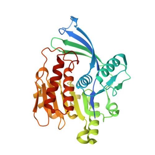
Ketohexokinase
A, B
313Homo sapiensMutation(s): 0 
Gene Names: KHK
EC: 2.7.1.3
UniProt & NIH Common Fund Data Resources
Find proteins for P50053 (Homo sapiens)
Explore P50053 
Go to UniProtKB:  P50053
PHAROS:  P50053
GTEx:  ENSG00000138030 
Entity Groups  
Sequence Clusters30% Identity50% Identity70% Identity90% Identity95% Identity100% Identity
UniProt GroupP50053
Sequence Annotations
Expand
  • Reference Sequence
Experimental Data & Validation

Experimental Data

  • Method: X-RAY DIFFRACTION
  • Resolution: 2.14 Å
  • R-Value Free: 0.254 
  • R-Value Work: 0.228 
  • R-Value Observed: 0.229 
  • Space Group: P 21 21 21
Unit Cell:
Length ( Å )Angle ( ˚ )
a = 83.36α = 90
b = 86.89β = 90
c = 137.91γ = 90
Software Package:
Software NamePurpose
BUSTERrefinement
PDB_EXTRACTdata extraction
XDSdata reduction
Aimlessdata scaling
BALBESphasing

Structure Validation

View Full Validation Report


Ligand Structure Quality Assessment 


Entry History & Funding Information

Deposition Data


Funding OrganizationLocationGrant Number
Not funded--

Revision History  (Full details and data files)

  • Version 1.0: 2023-09-27
    Type: Initial release
  • Version 1.1: 2023-10-18
    Changes: Database references