8OI6 | pdb_00008oi6

Cryo-EM structure of the undecorated barbed end of filamentous beta/gamma actin


Experimental Data Snapshot

  • Method: ELECTRON MICROSCOPY
  • Resolution: 3.59 Å
  • Aggregation State: PARTICLE 
  • Reconstruction Method: SINGLE PARTICLE 

Starting Model: experimental
View more details

wwPDB Validation 3D Report Full Report

Validation slider image for 8OI6

This is version 1.5 of the entry. See complete history

Literature

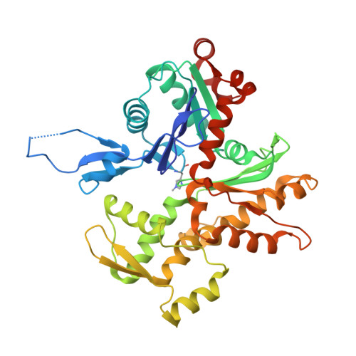
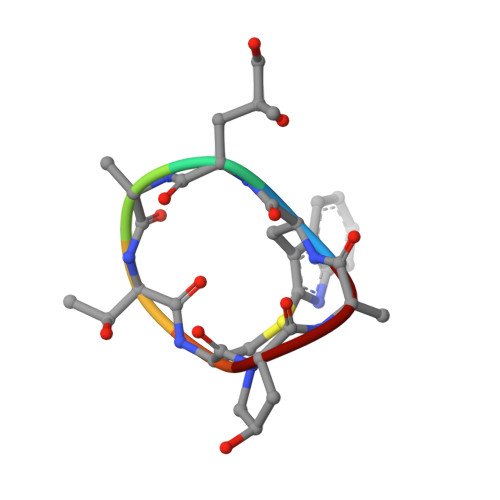
Molecular mechanisms of inorganic-phosphate release from the core and barbed end of actin filaments.

Oosterheert, W.Blanc, F.E.C.Roy, A.Belyy, A.Sanders, M.B.Hofnagel, O.Hummer, G.Bieling, P.Raunser, S.

(2023) Nat Struct Mol Biol 30: 1774-1785

  • DOI: https://doi.org/10.1038/s41594-023-01101-9
  • Primary Citation Related Structures: 
    8OI6, 8OI8, 8OID

  • PubMed Abstract: 

    The release of inorganic phosphate (P i ) from actin filaments constitutes a key step in their regulated turnover, which is fundamental to many cellular functions. The mechanisms underlying P i release from the core and barbed end of actin filaments remain unclear. Here, using human and bovine actin isoforms, we combine cryo-EM with molecular-dynamics simulations and in vitro reconstitution to demonstrate how actin releases P i through a 'molecular backdoor'. While constantly open at the barbed end, the backdoor is predominantly closed in filament-core subunits and opens only transiently through concerted amino acid rearrangements. This explains why P i escapes rapidly from the filament end but slowly from internal subunits. In a nemaline-myopathy-associated actin variant, the backdoor is predominantly open in filament-core subunits, resulting in accelerated P i release and filaments with drastically shortened ADP-P i caps. Our results provide the molecular basis for P i release from actin and exemplify how a disease-linked mutation distorts the nucleotide-state distribution and atomic structure of the filament.


  • Organizational Affiliation
    • Department of Structural Biochemistry, Max Planck Institute of Molecular Physiology, Dortmund, Germany.

Macromolecule Content 

  • Total Structure Weight: 171.42 kDa 
  • Atom Count: 11,573 
  • Modeled Residue Count: 1,467 
  • Deposited Residue Count: 1,521 
  • Unique protein chains: 2

Macromolecules

Find similar proteins by:|  3D Structure
Entity ID: 1
MoleculeChains  Sequence LengthOrganismDetailsImage
Actin, cytoplasmic 1
A, B, C, D
375Bos taurusMutation(s): 0 
EC: 3.6.4
UniProt
Find proteins for P60712 (Bos taurus)
Explore P60712 
Go to UniProtKB:  P60712
Entity Groups
Sequence Clusters30% Identity50% Identity70% Identity90% Identity95% Identity100% Identity
UniProt GroupP60712
Sequence Annotations
Expand
Reference Sequence
Find similar proteins by:  Sequence   |   3D Structure  
Entity ID: 2
MoleculeChains  Sequence LengthOrganismDetailsImage
PHALLOIDINE [auth H],
F [auth I],
G [auth J]
7Amanita phalloidesMutation(s): 0 

Small Molecules

Ligands 2 Unique
IDChains Name / Formula / InChI Key2D Diagram3D Interactions
ADP
(Subject of Investigation/LOI)

Query on ADP


Download:Ideal Coordinates CCD File
H [auth A],
J [auth B],
L [auth C],
N [auth D]
ADENOSINE-5'-DIPHOSPHATE
C10 H15 N5 O10 P2
XTWYTFMLZFPYCI-KQYNXXCUSA-N
MG
(Subject of Investigation/LOI)

Query on MG


Download:Ideal Coordinates CCD File
I [auth A],
K [auth B],
M [auth C],
O [auth D]
MAGNESIUM ION
Mg
JLVVSXFLKOJNIY-UHFFFAOYSA-N
Modified Residues  2 Unique
IDChains TypeFormula2D DiagramParent
HIC
Query on HIC
A, B, C, D
L-PEPTIDE LINKINGC7 H11 N3 O2HIS
DTH
Query on DTH
E [auth H],
F [auth I],
G [auth J]
D-PEPTIDE LINKINGC4 H9 N O3

--

EEP
Query on EEP
E [auth H],
F [auth I],
G [auth J]
L-PEPTIDE LINKINGC6 H13 N O4

--

HYP
Query on HYP
E [auth H],
F [auth I],
G [auth J]
L-PEPTIDE LINKINGC5 H9 N O3PRO

Biologically Interesting Molecules (External Reference) 

1 Unique

Experimental Data & Validation

Experimental Data

  • Method: ELECTRON MICROSCOPY
  • Resolution: 3.59 Å
  • Aggregation State: PARTICLE 
  • Reconstruction Method: SINGLE PARTICLE 
EM Software:
TaskSoftware PackageVersion
MODEL REFINEMENTPHENIX1.20.1-4487-000
RECONSTRUCTIONcryoSPARC3.3.2

Structure Validation

View Full Validation Report


Entry History 

& Funding Information

Deposition Data


Funding OrganizationLocationGrant Number
Max Planck SocietyGermany--
Alexander von Humboldt FoundationGermany--
European Research Council (ERC)European Union856118

Revision History  (Full details and data files)

  • Version 1.0: 2023-08-09
    Type: Initial release
  • Version 1.1: 2023-08-16
    Changes: Data collection, Database references
  • Version 1.2: 2023-09-27
    Changes: Database references, Refinement description
  • Version 1.3: 2023-10-04
    Changes: Database references
  • Version 1.4: 2023-11-15
    Changes: Data collection
  • Version 1.5: 2023-11-22
    Changes: Database references