8OFW | pdb_00008ofw

Crystal structure of the full-length dihydroorotate dehydrogenase from Mycobacterium tuberculosis


Experimental Data Snapshot

  • Method: X-RAY DIFFRACTION
  • Resolution: 3.80 Å
  • R-Value Free: 
    0.328 (Depositor), 0.331 (DCC) 
  • R-Value Work: 
    0.255 (Depositor), 0.257 (DCC) 

Starting Model: in silico
View more details

wwPDB Validation 3D Report Full Report

Validation slider image for 8OFW

Ligand Structure Quality Assessment 


This is version 1.0 of the entry. See complete history

Literature

Biochemical characterization of Mycobacterium tuberculosis dihydroorotate dehydrogenase and identification of a selective inhibitor.

Alberti, M.Sainas, S.Ronchi, E.Lolli, M.L.Boschi, D.Rizzi, M.Ferraris, D.M.Miggiano, R.

(2023) FEBS Lett 597: 2119-2132

  • DOI: https://doi.org/10.1002/1873-3468.14680
  • Primary Citation Related Structures: 
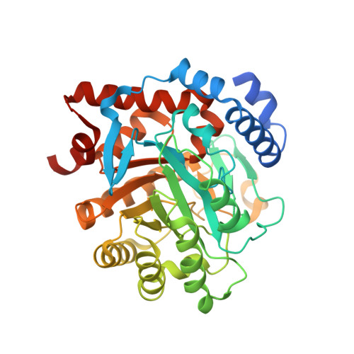
    8OFW

  • PubMed Abstract: 

    Mycobacterium tuberculosis (MTB) is the etiologic agent of tuberculosis (TB), an ancient disease which causes 1.5 million deaths worldwide. Dihydroorotate dehydrogenase (DHODH) is a key enzyme of the MTB de novo pyrimidine biosynthesis pathway, and it is essential for MTB growth in vitro, hence representing a promising drug target. We present: (i) the biochemical characterization of the full-length MTB DHODH, including the analysis of the kinetic parameters, and (ii) the previously unreleased crystal structure of the protein that allowed us to rationally screen our in-house chemical library and identify the first selective inhibitor of mycobacterial DHODH. The inhibitor has fluorescence properties, potentially instrumental to in cellulo imaging studies, and exhibits an IC 50 value of 43 μm, paving the way to hit-to-lead process.


  • Organizational Affiliation
    • Department of Pharmaceutical Sciences, University of Eastern Piedmont, Novara, Italy.

Macromolecule Content 

  • Total Structure Weight: 81.35 kDa 
  • Atom Count: 5,254 
  • Modeled Residue Count: 692 
  • Deposited Residue Count: 754 
  • Unique protein chains: 1

Macromolecules

Find similar proteins by:|  3D Structure
Entity ID: 1
MoleculeChains  Sequence LengthOrganismDetailsImage
Dihydroorotate dehydrogenase (quinone)
A, B
377Mycobacterium tuberculosisMutation(s): 0 
Gene Names: pyrDRv2139MTCY270.29c
EC: 1.3.5.2
UniProt
Find proteins for P9WHL1 (Mycobacterium tuberculosis (strain ATCC 25618 / H37Rv))
Explore P9WHL1 
Go to UniProtKB:  P9WHL1
Entity Groups
Sequence Clusters30% Identity50% Identity70% Identity90% Identity95% Identity100% Identity
UniProt GroupP9WHL1
Sequence Annotations
Expand
Reference Sequence

Experimental Data & Validation

Experimental Data

  • Method: X-RAY DIFFRACTION
  • Resolution: 3.80 Å
  • R-Value Free:  0.328 (Depositor), 0.331 (DCC) 
  • R-Value Work:  0.255 (Depositor), 0.257 (DCC) 
Space Group: P 21 21 21
Unit Cell:
Length ( Å )Angle ( ˚ )
a = 46.05α = 90
b = 85.215β = 90
c = 181.005γ = 90
Software Package:
Software NamePurpose
REFMACrefinement
XDSdata reduction
SCALAdata scaling
PHASERphasing

Structure Validation

View Full Validation Report


Ligand Structure Quality Assessment 


Entry History 

& Funding Information

Deposition Data


Funding OrganizationLocationGrant Number
Ministero dell Universita e della RicercaItaly--

Revision History  (Full details and data files)

  • Version 1.0: 2023-09-06
    Type: Initial release