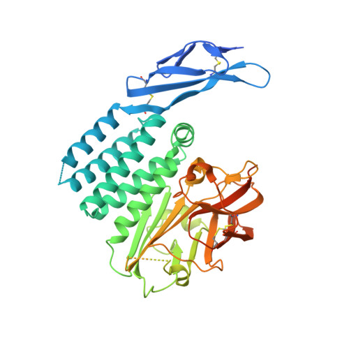
8OEK | pdb_00008oek

Crystal structure of the HormR-GAIN domains of adhesion GPCR ADGRB2 (BAI2) in the uncleaved state


Experimental Data Snapshot

  • Method: X-RAY DIFFRACTION
  • Resolution: 2.22 Å
  • R-Value Free: 
    0.287 (Depositor), 0.286 (DCC) 
  • R-Value Work: 
    0.229 (Depositor), 0.230 (DCC) 
  • R-Value Observed: 
    0.232 (Depositor) 

Starting Model: experimental
View more details

wwPDB Validation 3D Report Full Report

Validation slider image for 8OEK

Ligand Structure Quality Assessment 


This is version 1.2 of the entry. See complete history. 

Literature

Structural basis of GAIN domain autoproteolysis and cleavage-resistance in the adhesion G-protein coupled receptors.

Pohl, F., Seufert, F., Chung, Y.K., Schick, R., Kieslich, B., Schoneberg, T., Langenhan, T., Hildebrand, P.W., Strater, N.

(2026) Nat Commun 17

  • DOI: https://doi.org/10.1038/s41467-026-71225-1
  • Primary Citation Related Structures: 
    8OEK

  • PubMed Abstract: 

    The GAIN domain is a hallmark of adhesion G protein-coupled receptors (aGPCRs), as this extracellular domain contains an integral agonistic sequence (Stachel) that activates the receptor by binding to its 7-transmembrane helix (7TM) domain. Many aGPCRs undergo autoproteolytic cleavage at the GPCR proteolysis site (GPS), which has a canonical H-X-S/T sequence motif located within the GPCR autoproteolysis-inducing (GAIN) domain. Here we present the crystal structure of the Hormone Receptor (HormR) and GAIN domains of ADGRB2/BAI2. The protein was not cleaved at the GPS, despite possessing an HLS sequence. Through structural comparisons and molecular dynamics (MD) simulations, we identify determinants that contribute to autoproteolytic activity beyond the H-X-S/T motif. Specifically, we highlight a T-shaped π-π interaction between the histidine base of the H-X-S/T motif and a phenylalanine residue that is highly conserved in cleavage-competent aGPCRs. This interaction is critical for properly positioning the imidazole group of the histidine to deprotonate the alcohol nucleophile. Disruption of this interaction reduces autoproteolytic activity in the ADGRL1 and introduction of the phenylalanine restores cleavage competence in the otherwise non-cleavable ADGRB3 upon expression in HEK293 cells. In addition, the poorly conserved and flexible flap regions flanking the GPS also contribute to full autocleavage activity.


  • Organizational Affiliation: 
    • Institute of Bioanalytical Chemistry, Leipzig University, Leipzig, Germany.

Macromolecule Content 

  • Total Structure Weight: 49.74 kDa 
  • Atom Count: 2,754 
  • Modeled Residue Count: 344 
  • Deposited Residue Count: 449 
  • Unique protein chains: 1

Macromolecules

Find similar proteins by:|  3D Structure
Entity ID: 1
MoleculeChains  Sequence LengthOrganismDetailsImage
Adhesion G protein-coupled receptor B2449Homo sapiensMutation(s): 0 
Gene Names: ADGRB2, BAI2
UniProt & NIH Common Fund Data Resources
Find proteins for O60241 (Homo sapiens)
Explore O60241 
Go to UniProtKB:  O60241
PHAROS:  O60241
GTEx:  ENSG00000121753 
Entity Groups
Sequence Clusters30% Identity50% Identity70% Identity90% Identity95% Identity100% Identity
UniProt GroupO60241
Glycosylation
Glycosylation Sites: 1Go to GlyGen: O60241-1
Sequence Annotations
Expand
Reference Sequence

Small Molecules

Ligands 2 Unique
IDChains Name / Formula / InChI Key2D Diagram3D Interactions
NAG

Query on NAG


Download:Ideal Coordinates CCD File
B [auth A]2-acetamido-2-deoxy-beta-D-glucopyranose
C8 H15 N O6
OVRNDRQMDRJTHS-FMDGEEDCSA-N
CAC

Query on CAC


Download:Ideal Coordinates CCD File
C [auth A]CACODYLATE ION
C2 H6 As O2
OGGXGZAMXPVRFZ-UHFFFAOYSA-M

Experimental Data & Validation

Experimental Data

  • Method: X-RAY DIFFRACTION
  • Resolution: 2.22 Å
  • R-Value Free:  0.287 (Depositor), 0.286 (DCC) 
  • R-Value Work:  0.229 (Depositor), 0.230 (DCC) 
  • R-Value Observed: 0.232 (Depositor) 
Space Group: P 21 21 2
Unit Cell:
Length ( Å )Angle ( ˚ )
a = 84.864α = 90
b = 95.183β = 90
c = 49.465γ = 90
Software Package:
Software NamePurpose
PHENIXrefinement
XDSdata reduction
Aimlessdata scaling
STARANISOdata scaling

Structure Validation

View Full Validation Report


Ligand Structure Quality Assessment 


Entry History 

& Funding Information

Deposition Data


Funding OrganizationLocationGrant Number
German Research Foundation (DFG)Germany421152132

Revision History  (Full details and data files)

  • Version 1.0: 2023-05-31
    Type: Initial release
  • Version 1.1: 2024-11-06
    Changes: Data collection, Structure summary
  • Version 1.2: 2026-10-21
    Changes: Database references