8ODO | pdb_00008odo

Structure of human guanylylated RTCB in complex with Archease


Experimental Data Snapshot

  • Method: X-RAY DIFFRACTION
  • Resolution: 2.20 Å
  • R-Value Free: 
    0.218 (Depositor), 0.216 (DCC) 
  • R-Value Work: 
    0.193 (Depositor), 0.191 (DCC) 
  • R-Value Observed: 
    0.195 (Depositor) 

Starting Models: experimental
View more details

wwPDB Validation 3D Report Full Report

Validation slider image for 8ODO

Ligand Structure Quality Assessment 


This is version 1.2 of the entry. See complete history

Literature

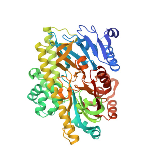
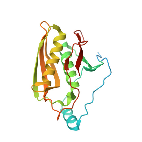
Structural and mechanistic insights into activation of the human RNA ligase RTCB by Archease.

Gerber, J.L.Morales Guzman, S.I.Worf, L.Hubbe, P.Kopp, J.Peschek, J.

(2024) Nat Commun 15: 2378-2378

  • DOI: https://doi.org/10.1038/s41467-024-46568-2
  • Primary Citation Related Structures: 
    8BTT, 8BTX, 8ODO, 8ODP

  • PubMed Abstract: 

    RNA ligases of the RTCB-type play an essential role in tRNA splicing, the unfolded protein response and RNA repair. RTCB is the catalytic subunit of the pentameric human tRNA ligase complex. RNA ligation by the tRNA ligase complex requires GTP-dependent activation of RTCB. This active site guanylylation reaction relies on the activation factor Archease. The mechanistic interplay between both proteins has remained unknown. Here, we report a biochemical and structural analysis of the human RTCB-Archease complex in the pre- and post-activation state. Archease reaches into the active site of RTCB and promotes the formation of a covalent RTCB-GMP intermediate through coordination of GTP and metal ions. During the activation reaction, Archease prevents futile RNA substrate binding to RTCB. Moreover, monomer structures of Archease and RTCB reveal additional states within the RNA ligation mechanism. Taken together, we present structural snapshots along the reaction cycle of the human tRNA ligase.


  • Organizational Affiliation
    • Heidelberg University, Biochemistry Center (BZH), Im Neuenheimer Feld 328, Heidelberg, Germany.

Macromolecule Content 

  • Total Structure Weight: 315.91 kDa 
  • Atom Count: 21,728 
  • Modeled Residue Count: 2,687 
  • Deposited Residue Count: 2,828 
  • Unique protein chains: 2

Macromolecules

Find similar proteins by:|  3D Structure
Entity ID: 1
MoleculeChains  Sequence LengthOrganismDetailsImage
RNA-splicing ligase RtcB homolog
A, C, E, G
507Homo sapiensMutation(s): 0 
Gene Names: RTCBC22orf28HSPC117
EC: 6.5.1.8
UniProt & NIH Common Fund Data Resources
Find proteins for Q9Y3I0 (Homo sapiens)
Explore Q9Y3I0 
Go to UniProtKB:  Q9Y3I0
PHAROS:  Q9Y3I0
GTEx:  ENSG00000100220 
Entity Groups
Sequence Clusters30% Identity50% Identity70% Identity90% Identity95% Identity100% Identity
UniProt GroupQ9Y3I0
Sequence Annotations
Expand
Reference Sequence
Find similar proteins by:|  3D Structure
Entity ID: 2
MoleculeChains  Sequence LengthOrganismDetailsImage
Protein archease
B, D, F, H
200Homo sapiensMutation(s): 0 
Gene Names: ZBTB8OS
EC: 2.7.7
UniProt & NIH Common Fund Data Resources
Find proteins for Q8IWT0 (Homo sapiens)
Explore Q8IWT0 
Go to UniProtKB:  Q8IWT0
PHAROS:  Q8IWT0
GTEx:  ENSG00000176261 
Entity Groups
Sequence Clusters30% Identity50% Identity70% Identity90% Identity95% Identity100% Identity
UniProt GroupQ8IWT0
Sequence Annotations
Expand
Reference Sequence

Small Molecules

Ligands 3 Unique
IDChains Name / Formula / InChI Key2D Diagram3D Interactions
5GP
(Subject of Investigation/LOI)

Query on 5GP


Download:Ideal Coordinates CCD File
I [auth A],
M [auth C],
Q [auth E],
U [auth G]
GUANOSINE-5'-MONOPHOSPHATE
C10 H14 N5 O8 P
RQFCJASXJCIDSX-UUOKFMHZSA-N
PO4

Query on PO4


Download:Ideal Coordinates CCD File
K [auth A],
O [auth C],
S [auth E],
W [auth G]
PHOSPHATE ION
O4 P
NBIIXXVUZAFLBC-UHFFFAOYSA-K
MN

Query on MN


Download:Ideal Coordinates CCD File
J [auth A]
L [auth B]
N [auth C]
P [auth D]
R [auth E]
J [auth A],
L [auth B],
N [auth C],
P [auth D],
R [auth E],
T [auth F],
V [auth G],
X [auth H]
MANGANESE (II) ION
Mn
WAEMQWOKJMHJLA-UHFFFAOYSA-N

Experimental Data & Validation

Experimental Data

  • Method: X-RAY DIFFRACTION
  • Resolution: 2.20 Å
  • R-Value Free:  0.218 (Depositor), 0.216 (DCC) 
  • R-Value Work:  0.193 (Depositor), 0.191 (DCC) 
  • R-Value Observed: 0.195 (Depositor) 
Space Group: P 1 21 1
Unit Cell:
Length ( Å )Angle ( ˚ )
a = 99.09α = 90
b = 125.547β = 108.51
c = 125.733γ = 90
Software Package:
Software NamePurpose
PHENIXrefinement
XDSdata reduction
Aimlessdata scaling
PHASERphasing

Structure Validation

View Full Validation Report


Ligand Structure Quality Assessment 


Entry History 

& Funding Information

Deposition Data


Funding OrganizationLocationGrant Number
German Research Foundation (DFG)Germany442512666

Revision History  (Full details and data files)

  • Version 1.0: 2024-03-27
    Type: Initial release
  • Version 1.1: 2024-04-17
    Changes: Database references
  • Version 1.2: 2024-11-06
    Changes: Structure summary