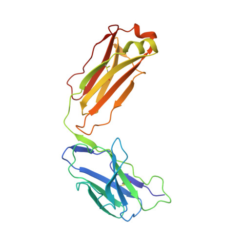
8KEI

Cryo-EM structure of NADPH oxidase 2 in complex with p22phox and EROS


Experimental Data Snapshot

  • Method: ELECTRON MICROSCOPY
  • Resolution: 3.56 Å
  • Aggregation State: PARTICLE 
  • Reconstruction Method: SINGLE PARTICLE 

wwPDB Validation   3D Report Full Report


This is version 1.2 of the entry. See complete history


Literature

Structural basis for EROS binding to human phagocyte NADPH oxidase NOX2.

Liang, S.Liu, A.Liu, Y.Wang, F.Zhou, Y.Long, Y.Wang, T.Liu, Z.Ren, R.Ye, R.D.

(2024) Proc Natl Acad Sci U S A 121: e2320388121-e2320388121

  • DOI: https://doi.org/10.1073/pnas.2320388121
  • Primary Citation of Related Structures:  
    8KEI

  • PubMed Abstract: 

    Essential for reactive oxygen species (EROS) protein is a recently identified molecular chaperone of NOX2 (gp91 phox ), the catalytic subunit of phagocyte NADPH oxidase. Deficiency in EROS is a recently identified cause for chronic granulomatous disease, a genetic disorder with recurrent bacterial and fungal infections. Here, we report a cryo-EM structure of the EROS-NOX2-p22 phox heterotrimeric complex at an overall resolution of 3.56Å. EROS and p22 phox are situated on the opposite sides of NOX2, and there is no direct contact between them. EROS associates with NOX2 through two antiparallel transmembrane (TM) α-helices and multiple β-strands that form hydrogen bonds with the cytoplasmic domain of NOX2. EROS binding induces a 79° upward bend of TM2 and a 48° backward rotation of the lower part of TM6 in NOX2, resulting in an increase in the distance between the two hemes and a shift of the binding site for flavin adenine dinucleotide (FAD). These conformational changes are expected to compromise superoxide production by NOX2, suggesting that the EROS-bound NOX2 is in a protected state against activation. Phorbol myristate acetate, an activator of NOX2 in vitro, is able to induce dissociation of NOX2 from EROS with concurrent increase in FAD binding and superoxide production in a transfected COS-7 model. In differentiated neutrophil-like HL-60, the majority of NOX2 on the cell surface is dissociated with EROS. Further studies are required to delineate how EROS dissociates from NOX2 during its transport to cell surface, which may be a potential mechanism for regulation of NOX2 activation.


  • Organizational Affiliation

    Kobilka Institute of Innovative Drug Discovery, School of Medicine, The Chinese University of Hong Kong, Shenzhen, Guangdong 518172, China.


Macromolecules
Find similar proteins by:  (by identity cutoff)  |  3D Structure
Entity ID: 1
MoleculeChains Sequence LengthOrganismDetailsImage
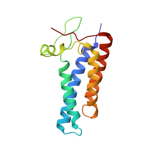
Cytochrome b-245 light chain133Homo sapiensMutation(s): 0 
Gene Names: CYBA
Membrane Entity: Yes 
UniProt & NIH Common Fund Data Resources
Find proteins for P13498 (Homo sapiens)
Explore P13498 
Go to UniProtKB:  P13498
PHAROS:  P13498
GTEx:  ENSG00000051523 
Entity Groups  
Sequence Clusters30% Identity50% Identity70% Identity90% Identity95% Identity100% Identity
UniProt GroupP13498
Sequence Annotations
Expand
  • Reference Sequence
Find similar proteins by:  (by identity cutoff)  |  3D Structure
Entity ID: 2
MoleculeChains Sequence LengthOrganismDetailsImage
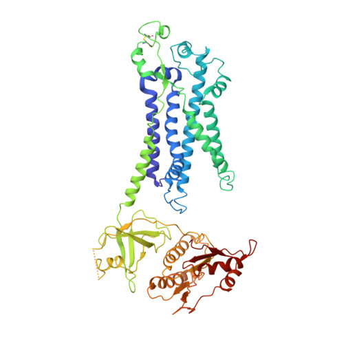
Cytochrome b-245 heavy chain565Homo sapiensMutation(s): 0 
Gene Names: CYBBNOX2
EC: 1
Membrane Entity: Yes 
UniProt & NIH Common Fund Data Resources
Find proteins for P04839 (Homo sapiens)
Explore P04839 
Go to UniProtKB:  P04839
PHAROS:  P04839
GTEx:  ENSG00000165168 
Entity Groups  
Sequence Clusters30% Identity50% Identity70% Identity90% Identity95% Identity100% Identity
UniProt GroupP04839
Glycosylation
Glycosylation Sites: 3Go to GlyGen: P04839-1
Sequence Annotations
Expand
  • Reference Sequence
Find similar proteins by:  (by identity cutoff)  |  3D Structure
Entity ID: 3
MoleculeChains Sequence LengthOrganismDetailsImage
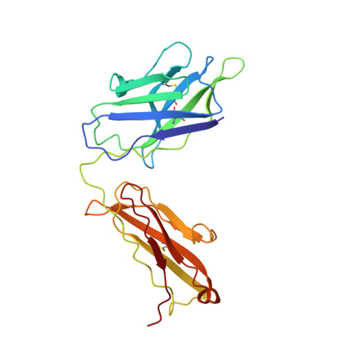
monoclonal antibody 7G5 heavy chain224Oryctolagus cuniculusMutation(s): 0 
Entity Groups  
Sequence Clusters30% Identity50% Identity70% Identity90% Identity95% Identity100% Identity
Sequence Annotations
Expand
  • Reference Sequence
Find similar proteins by:  (by identity cutoff)  |  3D Structure
Entity ID: 4
MoleculeChains Sequence LengthOrganismDetailsImage
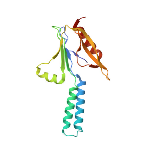
Cytochrome b-245 chaperone 1164Homo sapiensMutation(s): 0 
Gene Names: CYBC1C17orf62EROS
Membrane Entity: Yes 
UniProt & NIH Common Fund Data Resources
Find proteins for Q9BQA9 (Homo sapiens)
Explore Q9BQA9 
Go to UniProtKB:  Q9BQA9
PHAROS:  Q9BQA9
GTEx:  ENSG00000178927 
Entity Groups  
Sequence Clusters30% Identity50% Identity70% Identity90% Identity95% Identity100% Identity
UniProt GroupQ9BQA9
Sequence Annotations
Expand
  • Reference Sequence
Find similar proteins by:  (by identity cutoff)  |  3D Structure
Entity ID: 5
MoleculeChains Sequence LengthOrganismDetailsImage
monoclonal antibody 7G5 light chain217Oryctolagus cuniculusMutation(s): 0 
Entity Groups  
Sequence Clusters30% Identity50% Identity70% Identity90% Identity95% Identity100% Identity
Sequence Annotations
Expand
  • Reference Sequence
Oligosaccharides

Help

Entity ID: 6
MoleculeChains Length2D Diagram Glycosylation3D Interactions
beta-D-mannopyranose-(1-4)-2-acetamido-2-deoxy-beta-D-glucopyranose-(1-4)-2-acetamido-2-deoxy-beta-D-glucopyranose
F, G
3N-Glycosylation
Glycosylation Resources
GlyTouCan:  G15407YE
GlyCosmos:  G15407YE
GlyGen:  G15407YE
Small Molecules
Ligands 4 Unique
IDChains Name / Formula / InChI Key2D Diagram3D Interactions
LBN
Query on LBN

Download Ideal Coordinates CCD File 
L [auth B]1-palmitoyl-2-oleoyl-sn-glycero-3-phosphocholine
C42 H82 N O8 P
WTJKGGKOPKCXLL-VYOBOKEXSA-N
POV (Subject of Investigation/LOI)
Query on POV

Download Ideal Coordinates CCD File 
H [auth A](2S)-3-(hexadecanoyloxy)-2-[(9Z)-octadec-9-enoyloxy]propyl 2-(trimethylammonio)ethyl phosphate
C42 H82 N O8 P
WTJKGGKOPKCXLL-PFDVCBLKSA-N
HEM (Subject of Investigation/LOI)
Query on HEM

Download Ideal Coordinates CCD File 
I [auth B],
J [auth B]
PROTOPORPHYRIN IX CONTAINING FE
C34 H32 Fe N4 O4
KABFMIBPWCXCRK-RGGAHWMASA-L
NAG (Subject of Investigation/LOI)
Query on NAG

Download Ideal Coordinates CCD File 
K [auth B]2-acetamido-2-deoxy-beta-D-glucopyranose
C8 H15 N O6
OVRNDRQMDRJTHS-FMDGEEDCSA-N
Experimental Data & Validation

Experimental Data

  • Method: ELECTRON MICROSCOPY
  • Resolution: 3.56 Å
  • Aggregation State: PARTICLE 
  • Reconstruction Method: SINGLE PARTICLE 

Structure Validation

View Full Validation Report


Entry History & Funding Information

Deposition Data


Funding OrganizationLocationGrant Number
Not funded--

Revision History  (Full details and data files)

  • Version 1.0: 2024-05-22
    Type: Initial release
  • Version 1.1: 2024-11-20
    Changes: Data collection, Structure summary
  • Version 1.2: 2024-12-04
    Changes: Data collection, Database references