


Crystal structure of the Michaelis complex of trypsin with N-alpha-benzoyl-l-arginine ethyl ester
Akbar, Z., Ahmad, M.S.(2024) J Chin Chem Soc n/a
Experimental Data Snapshot
Starting Model: experimental
View more details
(2024) J Chin Chem Soc n/a
Entity ID: 1 | |||||
|---|---|---|---|---|---|
| Molecule | Chains | Sequence Length | Organism | Details | Image |
| Cationic trypsin | 223 | Bos taurus | Mutation(s): 0 EC: 3.4.21.4 | 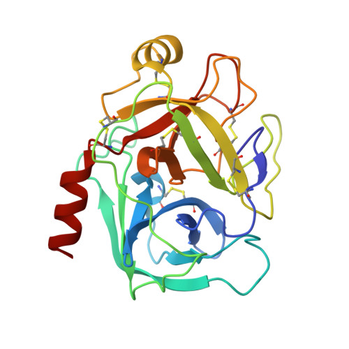 | |
UniProt | |||||
Entity Groups | |||||
| Sequence Clusters | 30% Identity50% Identity70% Identity90% Identity95% Identity100% Identity | ||||
| UniProt Group | P00760 | ||||
Sequence AnnotationsExpand | |||||
Reference Sequence | |||||
| Ligands 9 Unique | |||||
|---|---|---|---|---|---|
| ID | Chains | Name / Formula / InChI Key | 2D Diagram | 3D Interactions | |
| I4Q (Subject of Investigation/LOI) Download:Ideal Coordinates CCD File | B [auth A] | ethyl (2~{S})-2-benzamido-5-carbamimidamido-pentanoate C15 H22 N4 O3 YQDHCCVUYCIGSW-LBPRGKRZSA-N |  | ||
| JNR (Subject of Investigation/LOI) Download:Ideal Coordinates CCD File | C [auth A] | (2~{R})-2-benzamido-5-carbamimidamido-pentanoic acid C13 H18 N4 O3 RSYYQCDERUOEFI-SNVBAGLBSA-N |  | ||
| ARG Download:Ideal Coordinates CCD File | D [auth A] | ARGININE C6 H15 N4 O2 ODKSFYDXXFIFQN-BYPYZUCNSA-O |  | ||
| PEG Download:Ideal Coordinates CCD File | K [auth A] | DI(HYDROXYETHYL)ETHER C4 H10 O3 MTHSVFCYNBDYFN-UHFFFAOYSA-N |  | ||
| SO4 Download:Ideal Coordinates CCD File | F [auth A] | SULFATE ION O4 S QAOWNCQODCNURD-UHFFFAOYSA-L |  | ||
| GAI Download:Ideal Coordinates CCD File | E [auth A] | GUANIDINE C H5 N3 ZRALSGWEFCBTJO-UHFFFAOYSA-N |  | ||
| EOH Download:Ideal Coordinates CCD File | L [auth A], M [auth A] | ETHANOL C2 H6 O LFQSCWFLJHTTHZ-UHFFFAOYSA-N |  | ||
| CA Download:Ideal Coordinates CCD File | G [auth A] | CALCIUM ION Ca BHPQYMZQTOCNFJ-UHFFFAOYSA-N |  | ||
| CL Download:Ideal Coordinates CCD File | H [auth A], I [auth A], J [auth A] | CHLORIDE ION Cl VEXZGXHMUGYJMC-UHFFFAOYSA-M |  | ||
| Length ( Å ) | Angle ( ˚ ) |
|---|---|
| a = 131.116 | α = 90 |
| b = 131.116 | β = 90 |
| c = 71.424 | γ = 120 |
| Software Name | Purpose |
|---|---|
| REFMAC | refinement |
| PROTEUM PLUS | data reduction |
| Aimless | data scaling |
| MOLREP | phasing |
| Funding Organization | Location | Grant Number |
|---|---|---|
| Not funded | -- |


