8KCM | pdb_00008kcm

MmCPDII-DNA complex containing low-dosage, light induced repaired DNA.


Experimental Data Snapshot

  • Method: X-RAY DIFFRACTION
  • Resolution: 2.50 Å
  • R-Value Free: 
    0.259 (Depositor), 0.262 (DCC) 
  • R-Value Work: 
    0.218 (Depositor), 0.221 (DCC) 
  • R-Value Observed: 
    0.220 (Depositor) 

Starting Model: experimental
View more details

wwPDB Validation 3D Report Full Report

Validation slider image for 8KCM

Ligand Structure Quality Assessment 


This is version 1.2 of the entry. See complete history

Literature

Visualizing the DNA repair process by a photolyase at atomic resolution.

Maestre-Reyna, M.Wang, P.H.Nango, E.Hosokawa, Y.Saft, M.Furrer, A.Yang, C.H.Gusti Ngurah Putu, E.P.Wu, W.J.Emmerich, H.J.Caramello, N.Franz-Badur, S.Yang, C.Engilberge, S.Wranik, M.Glover, H.L.Weinert, T.Wu, H.Y.Lee, C.C.Huang, W.C.Huang, K.F.Chang, Y.K.Liao, J.H.Weng, J.H.Gad, W.Chang, C.W.Pang, A.H.Yang, K.C.Lin, W.T.Chang, Y.C.Gashi, D.Beale, E.Ozerov, D.Nass, K.Knopp, G.Johnson, P.J.M.Cirelli, C.Milne, C.Bacellar, C.Sugahara, M.Owada, S.Joti, Y.Yamashita, A.Tanaka, R.Tanaka, T.Luo, F.Tono, K.Zarzycka, W.Muller, P.Alahmad, M.A.Bezold, F.Fuchs, V.Gnau, P.Kiontke, S.Korf, L.Reithofer, V.Rosner, C.J.Seiler, E.M.Watad, M.Werel, L.Spadaccini, R.Yamamoto, J.Iwata, S.Zhong, D.Standfuss, J.Royant, A.Bessho, Y.Essen, L.O.Tsai, M.D.

(2023) Science 382: eadd7795-eadd7795

  • DOI: https://doi.org/10.1126/science.add7795
  • Primary Citation Related Structures: 
    7YC7, 7YCM, 7YCP, 7YCR, 7YD6, 7YD7, 7YD8, 7YDZ, 7YE0, 7YEB, 7YEC, 7YEE, 7YEI, 7YEJ, 7YEK, 7YEL, 7YEM, 8KCM

  • PubMed Abstract: 

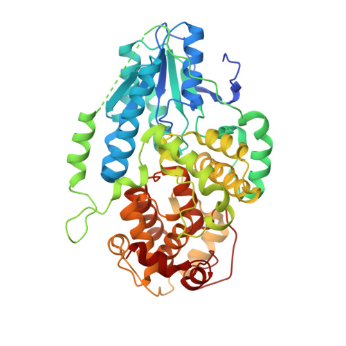
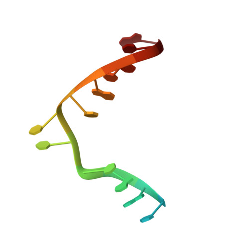
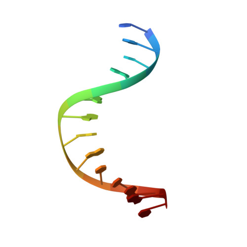
    Photolyases, a ubiquitous class of flavoproteins, use blue light to repair DNA photolesions. In this work, we determined the structural mechanism of the photolyase-catalyzed repair of a cyclobutane pyrimidine dimer (CPD) lesion using time-resolved serial femtosecond crystallography (TR-SFX). We obtained 18 snapshots that show time-dependent changes in four reaction loci. We used these results to create a movie that depicts the repair of CPD lesions in the picosecond-to-nanosecond range, followed by the recovery of the enzymatic moieties involved in catalysis, completing the formation of the fully reduced enzyme-product complex at 500 nanoseconds. Finally, back-flip intermediates of the thymine bases to reanneal the DNA were captured at 25 to 200 microseconds. Our data cover the complete molecular mechanism of a photolyase and, importantly, its chemistry and enzymatic catalysis at work across a wide timescale and at atomic resolution.


  • Organizational Affiliation
    • Institute of Biological Chemistry, Academia Sinica, 128 Academia Rd. Sec. 2, Nankang, Taipei 115, Taiwan.

Macromolecule Content 

  • Total Structure Weight: 129.87 kDa 
  • Atom Count: 8,706 
  • Modeled Residue Count: 931 
  • Deposited Residue Count: 1,020 
  • Unique protein chains: 1
  • Unique nucleic acid chains: 2

Macromolecules


Find similar proteins by:|  3D Structure
Entity ID: 1
MoleculeChains  Sequence LengthOrganismDetailsImage
Deoxyribodipyrimidine photo-lyase
A, B
482Methanosarcina mazei Go1Mutation(s): 1 
Gene Names: 
EC: 4.1.99.3
UniProt
Find proteins for Q8PYK9 (Methanosarcina mazei (strain ATCC BAA-159 / DSM 3647 / Goe1 / Go1 / JCM 11833 / OCM 88))
Explore Q8PYK9 
Go to UniProtKB:  Q8PYK9
Entity Groups
Sequence Clusters30% Identity50% Identity70% Identity90% Identity95% Identity100% Identity
UniProt GroupQ8PYK9
Sequence Annotations
Expand
Reference Sequence
Find similar nucleic acids by:  Sequence
Entity ID: 2
MoleculeChains LengthOrganismImage
Repaired DNA
C, E
14synthetic construct
Sequence Annotations
Expand
Reference Sequence
Find similar nucleic acids by:  Sequence
Entity ID: 3
MoleculeChains LengthOrganismImage
complementary oligonucleotide to the repaired DNA
D, F
14synthetic construct
Sequence Annotations
Expand
Reference Sequence

Small Molecules

Ligands 3 Unique
IDChains Name / Formula / InChI Key2D Diagram3D Interactions
FAD

Query on FAD


Download:Ideal Coordinates CCD File
G [auth A],
N [auth B]
FLAVIN-ADENINE DINUCLEOTIDE
C27 H33 N9 O15 P2
VWWQXMAJTJZDQX-UYBVJOGSSA-N
SO4

Query on SO4


Download:Ideal Coordinates CCD File
H [auth A]SULFATE ION
O4 S
QAOWNCQODCNURD-UHFFFAOYSA-L
GOL

Query on GOL


Download:Ideal Coordinates CCD File
I [auth A]
J [auth A]
K [auth A]
L [auth A]
M [auth A]
I [auth A],
J [auth A],
K [auth A],
L [auth A],
M [auth A],
O [auth B],
P [auth B],
Q [auth B],
R [auth B]
GLYCEROL
C3 H8 O3
PEDCQBHIVMGVHV-UHFFFAOYSA-N

Experimental Data & Validation

Experimental Data

  • Method: X-RAY DIFFRACTION
  • Resolution: 2.50 Å
  • R-Value Free:  0.259 (Depositor), 0.262 (DCC) 
  • R-Value Work:  0.218 (Depositor), 0.221 (DCC) 
  • R-Value Observed: 0.220 (Depositor) 
Space Group: P 21 21 21
Unit Cell:
Length ( Å )Angle ( ˚ )
a = 71.83α = 90
b = 115.04β = 90
c = 167.71γ = 90
Software Package:
Software NamePurpose
PHENIXrefinement
XDSdata reduction
XSCALEdata scaling
REFMACphasing

Structure Validation

View Full Validation Report


Ligand Structure Quality Assessment 


Entry History 

& Funding Information

Deposition Data


Funding OrganizationLocationGrant Number
Ministry of Science and Technology (MoST, Taiwan)TaiwanAS-KPQ-105-TPP
Ministry of Science and Technology (MoST, Taiwan)TaiwanAS-KPQ-109-TPP2
Japan Society for the Promotion of Science (JSPS)Japan16K01942

Revision History  (Full details and data files)

  • Version 1.0: 2023-11-22
    Type: Initial release
  • Version 1.1: 2023-12-13
    Changes: Database references
  • Version 1.2: 2024-03-13
    Changes: Source and taxonomy