8K78 | pdb_00008k78

Crystal structure of cMET kinase domain bound by TPX-0022


Experimental Data Snapshot

  • Method: X-RAY DIFFRACTION
  • Resolution: 2.67 Å
  • R-Value Free: 
    0.290 (Depositor), 0.290 (DCC) 
  • R-Value Work: 
    0.239 (Depositor), 0.240 (DCC) 
  • R-Value Observed: 
    0.244 (Depositor) 

wwPDB Validation 3D Report Full Report

Validation slider image for 8K78

Ligand Structure Quality Assessment 


This is version 1.0 of the entry. See complete history

Literature

Structural insight into the macrocyclic inhibitor TPX-0022 of c-Met and c-Src.

Qu, L.Lin, H.Dai, S.Guo, M.Chen, X.Jiang, L.Zhang, H.Li, M.Liang, X.Chen, Z.Wei, H.Chen, Y.

(2023) Comput Struct Biotechnol J 21: 5712-5718

  • DOI: https://doi.org/10.1016/j.csbj.2023.11.028
  • Primary Citation Related Structures: 
    8K78, 8K79

  • PubMed Abstract: 

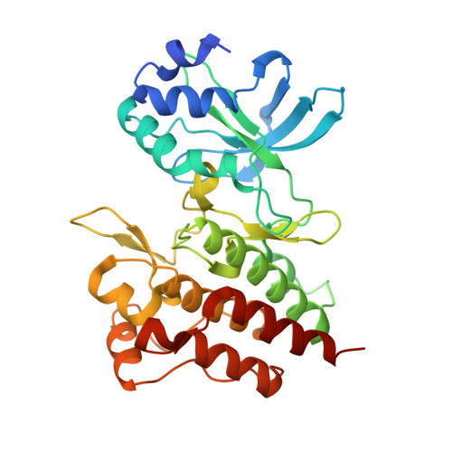
    c-Met has been an attractive target of prognostic and therapeutic studies in various cancers. TPX-0022 is a macrocyclic inhibitor of c-Met, c-Src and CSF1R kinases and is currently in phase I/II clinical trials in patients with advanced solid tumors harboring MET gene alterations. In this study, we determined the co-crystal structures of the c-Met/TPX-0022 and c-Src/TPX-0022 complexes to help elucidate the binding mechanism. TPX-0022 binds to the ATP pocket of c-Met and c-Src in a local minimum energy conformation and is stabilized by hydrophobic and hydrogen bond interactions. In addition, TPX-0022 exhibited potent activity against the resistance-relevant c-Met L1195F mutant and moderate activity against the c-Met G1163R, F1200I and Y1230H mutants but weak activity against the c-Met D1228N and Y1230C mutants. Overall, our study reveals the structural mechanism underlying the potency and selectivity of TPX-0022 and the ability to overcome acquire resistance mutations and provides insight into the development of selective c-Met macrocyclic inhibitors.


  • Organizational Affiliation
    • Department of Oncology, NHC Key Laboratory of Cancer Proteomics, State Local Joint Engineering Laboratory for Anticancer Drugs, Xiangya Hospital, Central South University, Changsha, Hunan 410008, China.

Macromolecule Content 

  • Total Structure Weight: 35.25 kDa 
  • Atom Count: 2,397 
  • Modeled Residue Count: 298 
  • Deposited Residue Count: 309 
  • Unique protein chains: 1

Macromolecules

Find similar proteins by:|  3D Structure
Entity ID: 1
MoleculeChains  Sequence LengthOrganismDetailsImage
Hepatocyte growth factor receptor309Homo sapiensMutation(s): 0 
Gene Names: MET
EC: 2.7.10.1
UniProt & NIH Common Fund Data Resources
Find proteins for P08581 (Homo sapiens)
Explore P08581 
Go to UniProtKB:  P08581
PHAROS:  P08581
GTEx:  ENSG00000105976 
Entity Groups
Sequence Clusters30% Identity50% Identity70% Identity90% Identity95% Identity100% Identity
UniProt GroupP08581
Sequence Annotations
Expand
Reference Sequence

Small Molecules

Ligands 1 Unique
IDChains Name / Formula / InChI Key2D Diagram3D Interactions
IYC
(Subject of Investigation/LOI)

Query on IYC


Download:Ideal Coordinates CCD File
B [auth A]Elzovantinib
C20 H20 F N7 O2
UUDPUQDMSHQSKH-NSHDSACASA-N
Binding Affinity Annotations 
IDSourceBinding Affinity
IYC BindingDB:  8K78 IC50: min: 0.14, max: 1.8 (nM) from 2 assay(s)

Experimental Data & Validation

Experimental Data

  • Method: X-RAY DIFFRACTION
  • Resolution: 2.67 Å
  • R-Value Free:  0.290 (Depositor), 0.290 (DCC) 
  • R-Value Work:  0.239 (Depositor), 0.240 (DCC) 
  • R-Value Observed: 0.244 (Depositor) 
Space Group: P 1 21 1
Unit Cell:
Length ( Å )Angle ( ˚ )
a = 45.054α = 90
b = 66.625β = 107
c = 55.294γ = 90
Software Package:
Software NamePurpose
PHENIXrefinement
PHASERphasing
XDSdata reduction
XDSdata scaling

Structure Validation

View Full Validation Report


Ligand Structure Quality Assessment 


Entry History 

& Funding Information

Deposition Data


Funding OrganizationLocationGrant Number
National Natural Science Foundation of China (NSFC)China--

Revision History  (Full details and data files)

  • Version 1.0: 2024-07-31
    Type: Initial release