8K6W | pdb_00008k6w

dUTPase of helicobacter pylori 26695


Experimental Data Snapshot

  • Method: X-RAY DIFFRACTION
  • Resolution: 2.50 Å
  • R-Value Free: 
    0.238 (Depositor), 0.233 (DCC) 
  • R-Value Work: 
    0.175 (Depositor), 0.182 (DCC) 

Starting Model: experimental
View more details

wwPDB Validation 3D Report Full Report

Validation slider image for 8K6W

This is version 1.1 of the entry. See complete history

Literature

Structural analysis of dUTPase from Helicobacter pylori reveals unusual activity for dATP.

Kumari, K.Aggarwal, S.Khan, F.M.Munde, M.Gourinath, S.

(2024) Int J Biol Macromol 282: 136937-136937

  • DOI: https://doi.org/10.1016/j.ijbiomac.2024.136937
  • Primary Citation Related Structures: 
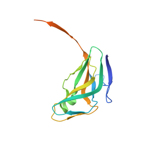
    8HRV, 8K6W

  • PubMed Abstract: 

    Helicobacter pylori deoxyuridine triphosphate nucleotidohydrolase (HpdUTPase) is a key enzyme in the synthesis of the thymidine nucleotide pathway. It catalyzes the hydrolysis of dUTP to dUMP and releases pyrophosphate. This enzyme has been shown to be essential in several pathogenic organisms. Here, we have determined the crystal structures of HpdUTPase in complex with α, β-imido dUTP (non-hydrolyzable substrate analog) and apo-state at resolution of 2 Å and 2.5 Å respectively. The flexible c terminal end of HpdUTPase which is not observed in apo-state structure and becomes ordered in the complex structure, suggesting its role in forming active site and substrate interaction. The Isothermal titration calorimetry (ITC) experiments reveal that hydrolysis of dUTP is an exothermic reaction with K m  = 35.0 ± 0.19 μM and the k cat  = 1.20 ± 0.19 s -1 . The ITC studies combined with MD simulations for all other nucleotides (dATP.dGTP, dCTP and dTTP) show that the active site of HpdUTPase strangely can also accommodate dATP. The structural comparison with the host (human) dUTPases reveals critical differences in substrate binding affinity of the active site of HpdUTPase. The detailed study suggests that the dATP binds in the active site of HpdUTPase making the number of preferable hydrogen bonds and shows activity with Km of 47 ± 2.4 μM.


  • Organizational Affiliation
    • Structural Biology Laboratory, School of Life Sciences, Jawaharlal Nehru University, New Delhi 110067, India.

Macromolecule Content 

  • Total Structure Weight: 50.17 kDa 
  • Atom Count: 3,004 
  • Modeled Residue Count: 388 
  • Deposited Residue Count: 456 
  • Unique protein chains: 1

Macromolecules

Find similar proteins by:|  3D Structure
Entity ID: 1
MoleculeChains  Sequence LengthOrganismDetailsImage
Deoxyuridine 5'-triphosphate nucleotidohydrolase
A, B, C
152Helicobacter pylori 26695Mutation(s): 0 
Gene Names: dutHP_0865
EC: 3.6.1.23
UniProt
Find proteins for O25536 (Helicobacter pylori (strain ATCC 700392 / 26695))
Explore O25536 
Go to UniProtKB:  O25536
Entity Groups
Sequence Clusters30% Identity50% Identity70% Identity90% Identity95% Identity100% Identity
UniProt GroupO25536
Sequence Annotations
Expand
Reference Sequence

Experimental Data & Validation

Experimental Data

  • Method: X-RAY DIFFRACTION
  • Resolution: 2.50 Å
  • R-Value Free:  0.238 (Depositor), 0.233 (DCC) 
  • R-Value Work:  0.175 (Depositor), 0.182 (DCC) 
Space Group: P 63
Unit Cell:
Length ( Å )Angle ( ˚ )
a = 102.36α = 90
b = 102.36β = 90
c = 78.37γ = 120
Software Package:
Software NamePurpose
REFMACrefinement
iMOSFLMdata reduction
Aimlessdata scaling
PHASERphasing

Structure Validation

View Full Validation Report


Entry History 

& Funding Information

Deposition Data


Funding OrganizationLocationGrant Number
Other governmentIndia--

Revision History  (Full details and data files)

  • Version 1.0: 2024-07-31
    Type: Initial release
  • Version 1.1: 2025-03-26
    Changes: Database references, Structure summary