8JY0

Crystal structure of RhoBAST complexed with TMR-DN


Experimental Data Snapshot

  • Method: X-RAY DIFFRACTION
  • Resolution: 2.75 Å
  • R-Value Free: 0.231 
  • R-Value Work: 0.202 
  • R-Value Observed: 0.203 

Starting Model: experimental
View more details

wwPDB Validation   3D Report Full Report


Ligand Structure Quality Assessment 


This is version 1.0 of the entry. See complete history


Literature

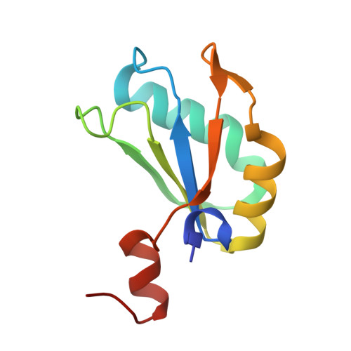
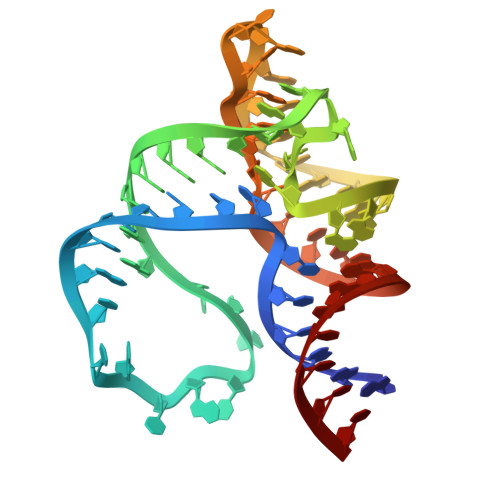
Structural mechanisms for binding and activation of a contact-quenched fluorophore by RhoBAST.

Zhang, Y.Xu, Z.Xiao, Y.Jiang, H.Zuo, X.Li, X.Fang, X.

(2024) Nat Commun 15: 4206-4206

  • DOI: https://doi.org/10.1038/s41467-024-48478-9
  • Primary Citation of Related Structures:  
    8JY0

  • PubMed Abstract: 

    The fluorescent light-up aptamer RhoBAST, which binds and activates the fluorophore-quencher conjugate tetramethylrhodamine-dinitroaniline with high affinity, super high brightness, remarkable photostability, and fast exchange kinetics, exhibits excellent performance in super-resolution RNA imaging. Here we determine the co-crystal structure of RhoBAST in complex with tetramethylrhodamine-dinitroaniline to elucidate the molecular basis for ligand binding and fluorescence activation. The structure exhibits an asymmetric "A"-like architecture for RhoBAST with a semi-open binding pocket harboring the xanthene of tetramethylrhodamine at the tip, while the dinitroaniline quencher stacks over the phenyl of tetramethylrhodamine instead of being fully released. Molecular dynamics simulations show highly heterogeneous conformational ensembles with the contact-but-unstacked fluorophore-quencher conformation for both free and bound tetramethylrhodamine-dinitroaniline being predominant. The simulations also show that, upon RNA binding, the fraction of xanthene-dinitroaniline stacked conformation significantly decreases in free tetramethylrhodamine-dinitroaniline. This highlights the importance of releasing dinitroaniline from xanthene tetramethylrhodamine to unquench the RhoBAST-tetramethylrhodamine-dinitroaniline complex. Using SAXS and ITC, we characterized the magnesium dependency of the folding and binding mode of RhoBAST in solution and indicated its strong structural robustness. The structures and binding modes of relevant fluorescent light-up aptamers are compared, providing mechanistic insights for rational design and optimization of this important fluorescent light-up aptamer-ligand system.


  • Organizational Affiliation

    Key Laboratory of RNA Science and Engineering, Institute of Biophysics Chinese Academy of Sciences, Beijing, China.


Macromolecules

Find similar proteins by:  (by identity cutoff)  |  3D Structure
Entity ID: 1
MoleculeChains Sequence LengthOrganismDetailsImage
U1 small nuclear ribonucleoprotein AA,
C,
D [auth E]
102Homo sapiensMutation(s): 2 
Gene Names: SNRPA
UniProt & NIH Common Fund Data Resources
Find proteins for P09012 (Homo sapiens)
Explore P09012 
Go to UniProtKB:  P09012
PHAROS:  P09012
GTEx:  ENSG00000077312 
Entity Groups  
Sequence Clusters30% Identity50% Identity70% Identity90% Identity95% Identity100% Identity
UniProt GroupP09012
Sequence Annotations
Expand
  • Reference Sequence
Find similar nucleic acids by:  (by identity cutoff)  |  3D Structure
Entity ID: 2
MoleculeChains LengthOrganismImage
RhoBASTB,
E [auth D],
F
64artificial sequences
Sequence Annotations
Expand
  • Reference Sequence
Small Molecules
Ligands 5 Unique
IDChains Name / Formula / InChI Key2D Diagram3D Interactions
GDP
Query on GDP

Download Ideal Coordinates CCD File 
I [auth B],
P [auth D],
R [auth F]
GUANOSINE-5'-DIPHOSPHATE
C10 H15 N5 O11 P2
QGWNDRXFNXRZMB-UUOKFMHZSA-N
V8C (Subject of Investigation/LOI)
Query on V8C

Download Ideal Coordinates CCD File 
K [auth B],
O [auth D],
Q [auth F]
5-aminocarbonyl-2-[3-(dimethylamino)-6-dimethylazaniumylidene-xanthen-9-yl]benzoate
C25 H23 N3 O4
TZKHAYIVKGBSEZ-UHFFFAOYSA-N
V7U (Subject of Investigation/LOI)
Query on V7U

Download Ideal Coordinates CCD File 
L [auth B]2,4-dinitroaniline
C6 H5 N3 O4
LXQOQPGNCGEELI-UHFFFAOYSA-N
PGE (Subject of Investigation/LOI)
Query on PGE

Download Ideal Coordinates CCD File 
J [auth B]TRIETHYLENE GLYCOL
C6 H14 O4
ZIBGPFATKBEMQZ-UHFFFAOYSA-N
MG
Query on MG

Download Ideal Coordinates CCD File 
G [auth B],
H [auth B],
M [auth D],
N [auth D]
MAGNESIUM ION
Mg
JLVVSXFLKOJNIY-UHFFFAOYSA-N
Experimental Data & Validation

Experimental Data

  • Method: X-RAY DIFFRACTION
  • Resolution: 2.75 Å
  • R-Value Free: 0.231 
  • R-Value Work: 0.202 
  • R-Value Observed: 0.203 
  • Space Group: I 21 21 21
Unit Cell:
Length ( Å )Angle ( ˚ )
a = 108.531α = 90
b = 158.936β = 90
c = 182.424γ = 90
Software Package:
Software NamePurpose
PHENIXrefinement
HKL-2000data scaling
HKL-2000data reduction
PHENIXphasing

Structure Validation

View Full Validation Report


Ligand Structure Quality Assessment 


Entry History & Funding Information

Deposition Data


Funding OrganizationLocationGrant Number
National Natural Science Foundation of China (NSFC)China--

Revision History  (Full details and data files)

  • Version 1.0: 2024-05-29
    Type: Initial release