8JVV

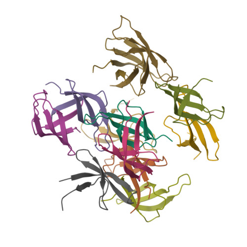
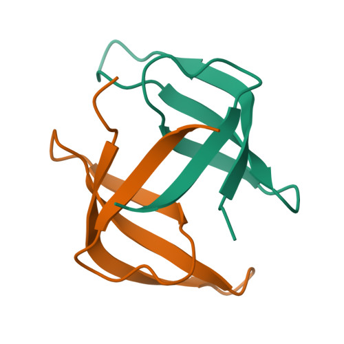
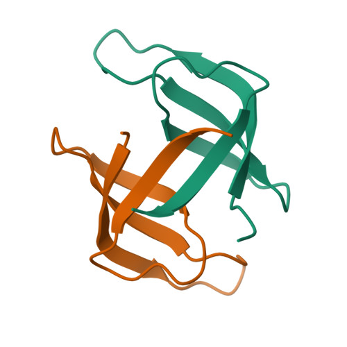
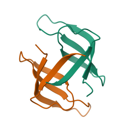
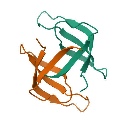
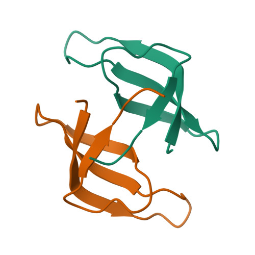
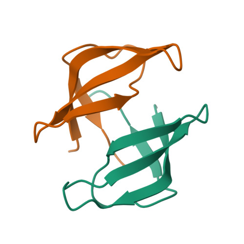
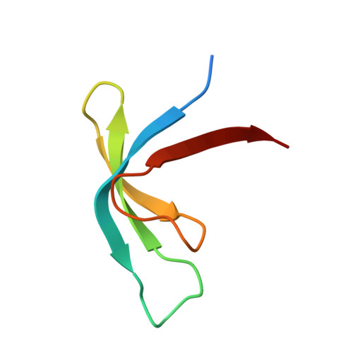
Crystal structure of the chimeric protein mkaL2_v1.1


Experimental Data Snapshot

  • Method: X-RAY DIFFRACTION
  • Resolution: 2.30 Å
  • R-Value Free: 0.252 
  • R-Value Work: 0.230 
  • R-Value Observed: 0.232 

Starting Model: experimental
View more details

wwPDB Validation   3D Report Full Report


This is version 1.1 of the entry. See complete history


Literature

An ancestral fold reveals the evolutionary link between RNA polymerase and ribosomal proteins.

Yagi, S.Tagami, S.

(2024) Nat Commun 15: 5938-5938

  • DOI: https://doi.org/10.1038/s41467-024-50013-9
  • Primary Citation of Related Structures:  
    8JVN, 8JVO, 8JVP, 8JVQ, 8JVR, 8JVS, 8JVT, 8JVU, 8JVV, 8JVW, 8JVX, 8JVY, 8JVZ

  • PubMed Abstract: 

    Numerous molecular machines are required to drive the central dogma of molecular biology. However, the means by which these numerous proteins emerged in the early evolutionary stage of life remains enigmatic. Many of them possess small β-barrel folds with different topologies, represented by double-psi β-barrels (DPBBs) conserved in DNA and RNA polymerases, and similar but topologically distinct six-stranded β-barrel RIFT or five-stranded β-barrel folds such as OB and SH3 in ribosomal proteins. Here, we discover that the previously reconstructed ancient DPBB sequence could also adopt a β-barrel fold named Double-Zeta β-barrel (DZBB), as a metamorphic protein. The DZBB fold is not found in any modern protein, although its structure shares similarities with RIFT and OB. Indeed, DZBB could be transformed into them through simple engineering experiments. Furthermore, the OB designs could be further converted into SH3 by circular-permutation as previously predicted. These results indicate that these β-barrels diversified quickly from a common ancestor at the beginning of the central dogma evolution.


  • Organizational Affiliation

    RIKEN Center for Biosystems Dynamics Research, 1-7-22 Suehiro-cho, Tsurumi-ku, Yokohama, Kanagawa, 230-0045, Japan. sota.yagi@aoni.waseda.jp.


Macromolecules
Find similar proteins by:  (by identity cutoff)  |  3D Structure
Entity ID: 1
MoleculeChains Sequence LengthOrganismDetailsImage
mkaL2_v1.1
A, B, C, D, E
A, B, C, D, E, F, G, H, I, J, K, L
55synthetic constructMutation(s): 0 
Entity Groups  
Sequence Clusters30% Identity50% Identity70% Identity90% Identity95% Identity100% Identity
Sequence Annotations
Expand
  • Reference Sequence
Experimental Data & Validation

Experimental Data

  • Method: X-RAY DIFFRACTION
  • Resolution: 2.30 Å
  • R-Value Free: 0.252 
  • R-Value Work: 0.230 
  • R-Value Observed: 0.232 
  • Space Group: P 1 21 1
Unit Cell:
Length ( Å )Angle ( ˚ )
a = 56.304α = 90
b = 95.351β = 114.1
c = 64.46γ = 90
Software Package:
Software NamePurpose
PHENIXrefinement
XDSdata reduction
XDSdata scaling
PHENIXphasing

Structure Validation

View Full Validation Report


Entry History & Funding Information

Deposition Data


Funding OrganizationLocationGrant Number
Japan Society for the Promotion of Science (JSPS)Japan18H01328
Japan Society for the Promotion of Science (JSPS)Japan20K15854
Japan Society for the Promotion of Science (JSPS)Japan22H01346

Revision History  (Full details and data files)

  • Version 1.0: 2024-07-03
    Type: Initial release
  • Version 1.1: 2024-08-07
    Changes: Database references