


Crystal Structure of Bel-1 E166A Mutant
Dhankhar, K., Hazra, S.To be published.
Experimental Data Snapshot
Starting Model: experimental
View more details
Entity ID: 1 | |||||
|---|---|---|---|---|---|
| Molecule | Chains | Sequence Length | Organism | Details | Image |
| Beta-lactamase | 263 | Pseudomonas aeruginosa | Mutation(s): 1 Gene Names: bla(BEL1) EC: 3.5.2.6 | 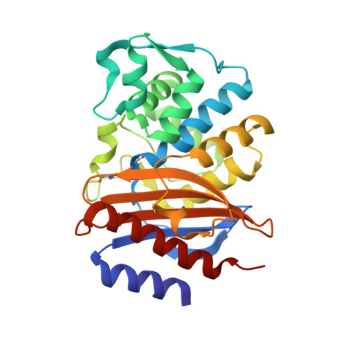 | |
UniProt | |||||
Entity Groups | |||||
| Sequence Clusters | 30% Identity50% Identity70% Identity90% Identity95% Identity100% Identity | ||||
| UniProt Group | Q3SAW3 | ||||
Sequence AnnotationsExpand | |||||
Reference Sequence | |||||
| Ligands 3 Unique | |||||
|---|---|---|---|---|---|
| ID | Chains | Name / Formula / InChI Key | 2D Diagram | 3D Interactions | |
| MES (Subject of Investigation/LOI) Download:Ideal Coordinates CCD File | C [auth A] | 2-(N-MORPHOLINO)-ETHANESULFONIC ACID C6 H13 N O4 S SXGZJKUKBWWHRA-UHFFFAOYSA-N |  | ||
| PO4 (Subject of Investigation/LOI) Download:Ideal Coordinates CCD File | E [auth A] | PHOSPHATE ION O4 P NBIIXXVUZAFLBC-UHFFFAOYSA-K |  | ||
| BR (Subject of Investigation/LOI) Download:Ideal Coordinates CCD File | D [auth A] | BROMIDE ION Br CPELXLSAUQHCOX-UHFFFAOYSA-M |  | ||
| Length ( Å ) | Angle ( ˚ ) |
|---|---|
| a = 123.498 | α = 90 |
| b = 123.498 | β = 90 |
| c = 70.644 | γ = 120 |
| Software Name | Purpose |
|---|---|
| REFMAC | refinement |
| CrysalisPro | data reduction |
| Aimless | data scaling |
| MOLREP | phasing |
| Funding Organization | Location | Grant Number |
|---|---|---|
| Other private | India | -- |


