


Crystal structure of Rv2186c in Mycobacterium tuberculosis
Yan, L., Zhang, R.To be published.
Experimental Data Snapshot
Starting Model: in silico
View more details
wwPDB Validation 3D Report Full Report

Entity ID: 1 | |||||
|---|---|---|---|---|---|
| Molecule | Chains | Sequence Length | Organism | Details | Image |
| Polyketide cyclase / dehydrase and lipid transport | A [auth B], B [auth A] | 129 | Mycobacterium tuberculosis | Mutation(s): 0 Gene Names: Rv2186c | 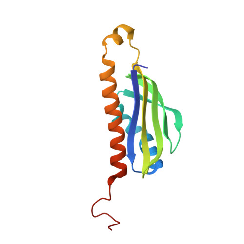 |
UniProt | |||||
Entity Groups | |||||
| Sequence Clusters | 30% Identity50% Identity70% Identity90% Identity95% Identity100% Identity | ||||
| UniProt Group | O53520 | ||||
Sequence AnnotationsExpand | |||||
Reference Sequence | |||||
| Length ( Å ) | Angle ( ˚ ) |
|---|---|
| a = 106.477 | α = 90 |
| b = 28.254 | β = 100.771 |
| c = 86.779 | γ = 90 |
| Software Name | Purpose |
|---|---|
| REFMAC | refinement |
| HKL-2000 | data reduction |
| HKL-2000 | data scaling |
| MOLREP | phasing |
| Funding Organization | Location | Grant Number |
|---|---|---|
| Ministry of Science and Technology (MoST, China) | China | 2017YFA0505900 |
| Other government | China | 2020YJ0209 |


